Origination of REiLI
In Japanese, the word “REiLI” connotes intelligent and resourceful.
REiLI is our AI technology brand which create new value by collaboration of AI with human and other AI.
By combining its existing image-processing technologies and natural language processing capabilities with AI, we are striving to create an open platform with wide-ranging value that provides robust support for diagnostic imaging and clinical workflows.
By combining its existing image-processing technologies and natural language processing capabilities with AI, we are striving to create an open platform with wide-ranging value that provides robust support for diagnostic imaging and clinical workflows.
Concept movie
I am REiLI.
Ever since I was born I have been involved in the medical field.
I know you never see me
but I am around when you’re helping, saving, and bettering people’s lives.
My mission is to be as good as I can be to help and better the work you do.
I am not one to improvise nor think outside the box but you can count on me to be immaculate in all that I do.
I maybe overly detailed and precise
but I hope that you can appreciate that meticulous side of me.
I never sleep.
So I am good to go day or night and deal with a whole lot of information.
There is so much to life
thus my mind is open
to learn from all minds
to consolidate that knowledge
to share with all.
I know you don’t see me but I exist in what you do.
My wish is to better the world through your patient care.
I am Here.
Invisibly.
REiLI”
History
1999
1983
Launched the world’s first digital radiography system
1996
Launched patented image intelligence algorithms in the consumer photo market place

1999
Released the industry’s first web-based Radiology PACS
2008
Launched 3D simulator for organ recognition/Resection
2015
Launched enterprise viewer in JAPAN which integrates various type of medical data into one system and optimizes medical professional’s workflow
2018
Launched the REiLI artificial intelligence platform and deep learning engines brand
2019
Released viewer which is optimized for AI usage in radiology department workflow(Provided only in Japan)
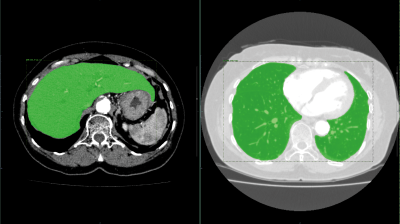
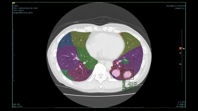
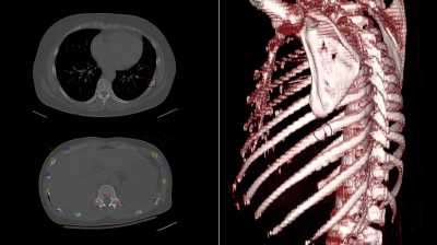
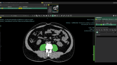
Organ segmentation and labelling technology
These technologies automatically extract organs such as liver, kidney, and spleen from CT images. They also extract cervical, thoracic, lumbar vertebrae and label them automatically.
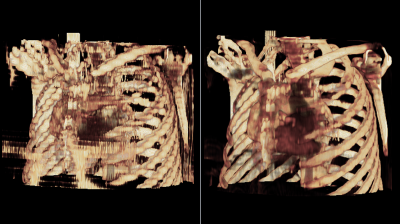
Thin slice virtual generation technology
The technology virtually generate thin slices from thick slices. It can be applied to the whole body, useful for utilizing past data. It brings high visibility in the VR display and reconstructed sagittal/coronal images.
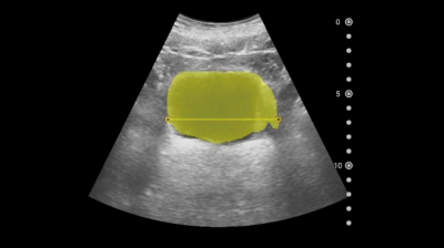
Bladder volume measurement
We are developing a technology to recognize urinary bladder area and measure the volume automatically.
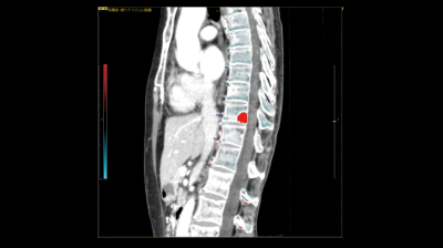
Bone Temporal Subtraction
This technology visualizes the bone density temporal difference by performing image registration between the past and current image of the same patient.
The increase and decrease in CT value will be highlighted.
2020
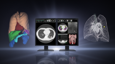
Lung labelling・Lung nodule CAD・Lung nodule characterization analysis
These technologies detect lung nodules from chest CT images, analyze multiple properties of nodules, and support populating radiology reports.
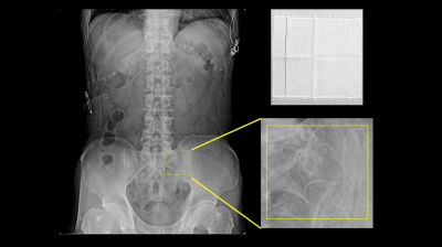
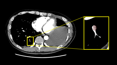
Retained object detection
This AI technology detects foreign objects in X-ray images. This technology can be applied to the detection of surgical objects such as gauze and reduce the burden on doctors to check the surgical objects held at each surgical procedure.
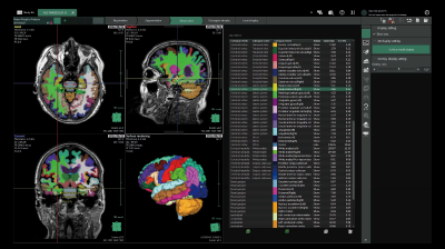
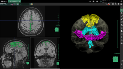
Brain Segmentation
AI technology to segment and quantify the volume of each brain regions.
This technology can be used for pre-surgery simulation or calculation of the atrophy rate for each region between past and current exams.
This technology can be used for pre-surgery simulation or calculation of the atrophy rate for each region between past and current exams.
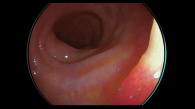
Detection Assist technology for colonic polyps
This technology assists real time detection and characterization of colonic polyps from colonoscopy images with AI software.
2021
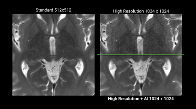
Denoising technique utilizing AI technology
We are developing the AI image reconstruction technology that removes noise components of image by preventing the structure and the contrast from degrading by a high speed or a high resolution MR scanning.
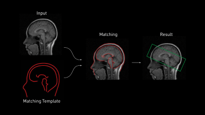
Improve workflow with AI
This technique extracts the target area from the image.
Positioning during imaging is performed automatically.
Unnecessary tissue is automatically removed before creating a 3D image for diagnosis.
It is a technology that supports the automation of inspections.
Positioning during imaging is performed automatically.
Unnecessary tissue is automatically removed before creating a 3D image for diagnosis.
It is a technology that supports the automation of inspections.
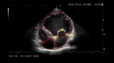
Automatic view recognition,Automatic measurement
The technology enables automatic recognition of left ventricle, left atrium and right atrium wall in addition to view recognition of the heart, and automatic measurements realize autonomous measurements of ejection fraction and volumes.
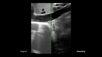
Noise reduction technology with AI
This technology uses AI to distinguish between echo signals and noises, and extracts the signals necessary for diagnosis.
Expantion of AI-CAD
The technology which prepares environment on the modality exapnds the opportuities of AI-CAD applications and improve workflow in wide range of use cases.
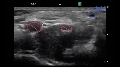
blood vessel detection technology
This technology assists real time detection and distinction between an artery and a vein, as well as measurement of diameter and depth of the vessel from ultrasound images. This will streamline Point Of Care UltraSound exams.
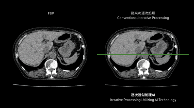
Iterative processing utilizing AI technology
The technology controls image quality based on a statistical model, an object model and a physical model using iterative processing.
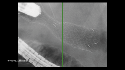
Super resolution processing
This flitering technology with AI suppresses aliasing noises which occur in extended images.
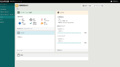
Cloud AI Platform
Released Cloud AI platform service (Provided only in Japan) Japan
Rib fracture CAD
AI technology to detect a suspcious rib fracture from CT images. This technology will assist prevention of overlooking of subtle rib fracture.
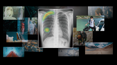
COVID CAD
AI technology to identify suspicious region with COVID-19 related findings from CT images. This technology will help doctors diagnose efficiently.
2022
SYNAPSE Creative Space
Released cloud AI development platform service to assist healthcare professionals and enginieers in develpoing AI technology.
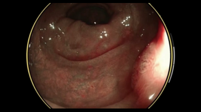
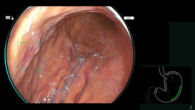
Detection technology for gastric neoplastic lesions and suspected esophageal squamous cell carcinoma
The technology utilizes AI to analyze upper gastrointestinal endoscopic images, recognizing areas suspected of being gastric neoplastic lesions or esophageal squamous cell carcinoma and detecting them in real time.
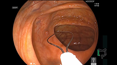
Endoscopy report creation support technology
In lower gastrointestinal endoscopy, AI technology is used to automatically recognize the insertion and removal of the scope from the body, as well as the insertion of surgical instruments, through software.
This is expected to support the recording of examination times and information on specimen collection (e.g., polyps), thereby reducing the burden on physicians in preparing reports.
This is expected to support the recording of examination times and information on specimen collection (e.g., polyps), thereby reducing the burden on physicians in preparing reports.
2023
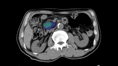
AI technology for abdominal CT
Technology that displays areas of high and low absorption in the liver, kidneys, and spleen compared to surrounding tissues.
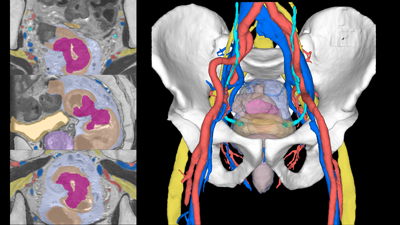
Pelvic segmentation technology
The technology for segmenting the rectum and surrounding organs from MRI images,which is expected to be useful for surgical simulation in the lower digestive tract area.
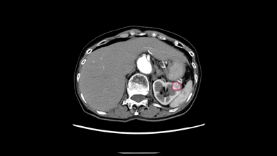
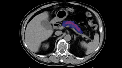
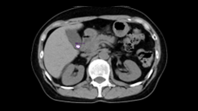
Pancreatic cancer detection technology
The technology that utilizes AI to help detect findings suggestive of pancreatic cancer from abdominal CT images.
2024
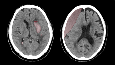
CT technology for diagnosing subarachnoid hemorrhage
We have developed AI technology to identify areas suspected of cerebral hemorrhage or cerebral infarction in CT images of the head. This technology is expected to aid in the diagnosis of stroke by helping to evaluate bleeding and ischemia in the brain.
Cerebrospinal fluid analysis technology in MRI images
We have developed AI technology to extract each region of the cerebrospinal fluid cavity from MRI images.
The aim is to improve diagnostic accuracy for Hakim’s disease (iNPH), a form of dementia that is important to detect early for treatment.
This technology is expected to improve the objectivity and accuracy of diagnosis by enabling AI to efficiently analyze regions associated with key findings (DESH) and support differentiation from brain atrophy.
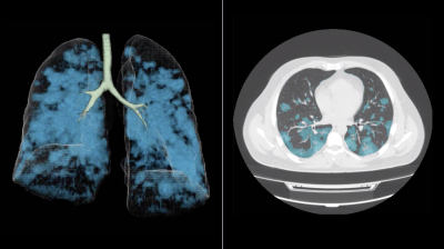
Pulmonary artery absorption enhancement technique
The technology that displays areas of low absorption in the pulmonary artery compared to surrounding tissues.
This is expected to aid in the diagnosis of pulmonary embolism.
This is expected to aid in the diagnosis of pulmonary embolism.
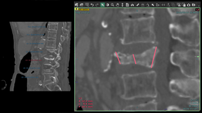
Automatic vertebral body height measurement technology
The technology that automatically measures the height of each vertebral body and displays the results classified according to user-defined thresholds.
This technology is expected to aid in the diagnosis of vertebral fractures.
This technology is expected to aid in the diagnosis of vertebral fractures.
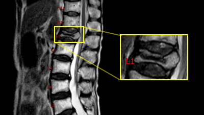
MRI labeling technology
The technique for labeling vertebral numbers on MRI images.
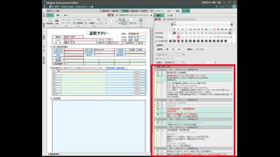
Medical record summary support technology
When creating documents such as discharge summaries, we developed a technology that extracts necessary text from medical records to assist.
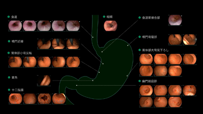
Endoscopy report creation support technology
In upper gastrointestinal endoscopy, AI technology is used to automatically recognize the location of images taken by doctors.
By extracting appropriate images for each location and automatically recording them in a report, it is expected that the burden of report creation for doctors will be reduced.
By extracting appropriate images for each location and automatically recording them in a report, it is expected that the burden of report creation for doctors will be reduced.
2025
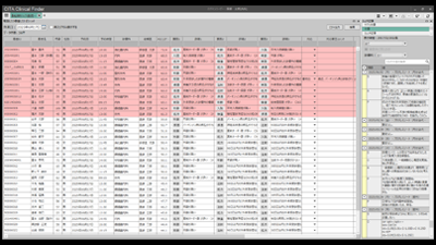
AI technology for predicting fall risk in outpatients
Based on clinical data accumulated in “CITA Clinical Finder,”
we have developed technology that predicts the risk of falls among outpatients, provides risk scores, and
identifies factors contributing to AI predictions.
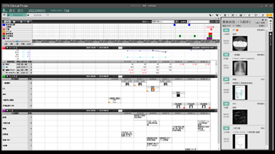
AI technology for medical data reference support
We have developed technology that predicts and lists medical data that is likely to be viewed based on patient information accumulated in “CITA Clinical Finder” and the viewing status of users, thereby supporting the reference of medical data.
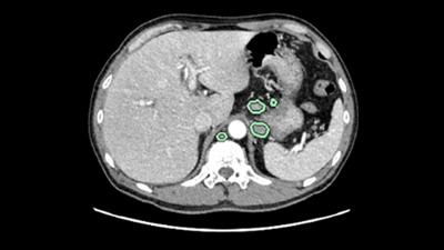
AI technology for abdominal CT
The technology that highlights areas of high/low absorption in the adrenal glands, pancreas, gallbladder, and lymph nodes compared to surrounding tissues.
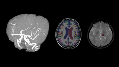
AI technology for head MRI
High signal/low signal region extraction technology, brain region labeling technology, brain extraction technology.
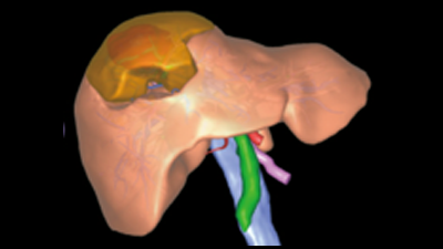
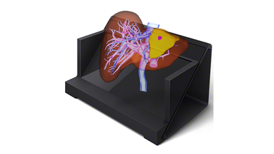
Liver deformation technology
Observe the liver and surrounding organs while deforming them, enabling estimation of the position of blood vessels during surgical detachment.
Naked-eye stereoscopic technology
Display 3D images created in the target application on a spatial reproduction display, and operate them in synchronization with our 3D system.
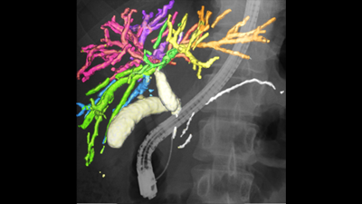
AI technology for the gallbladder and pancreas
Displays the complex structure of the gallbladder and pancreas in three dimensions, supporting preoperative simulation of organ relationships and other factors.
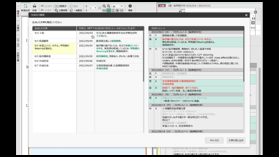
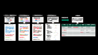
Natural language processing technology “AI for structuring medical reports”
Fujifilm has developed “Reading Report Structuring AI,” a unique natural language processing technology that structures reading reports created by radiologists for medical images captured by CT, MRI, and other imaging devices.
By structuring reading reports written based on the specialized knowledge of radiologists, it is possible to efficiently database and utilize reading reports that include medical jargon and expressions unique to physicians.
By structuring reading reports written based on the specialized knowledge of radiologists, it is possible to efficiently database and utilize reading reports that include medical jargon and expressions unique to physicians.
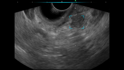
Ultrasound endoscopic diagnostic support technology for the pancreatic region
We have developed ultrasound endoscopy diagnostic support software that detects areas suspected of pancreatic solid lesions in real time during ultrasound endoscopy examinations, thereby supporting the early detection of pancreatic cancer. By analyzing ultrasound endoscopy images, the software detects areas where the pancreas is presumed to exist and areas suspected of pancreatic solid lesions in real time, and displays the results on the monitor’s ultrasound endoscopy image.By alerting the operator, the software assists in detecting pancreatic solid lesions.
Summary creation support technology
The technology utilizes a large-scale language model to automatically generate and present drafts of “summaries,” which are documents that comprehensively describe patient information such as test results and treatment history.