Interstitial lung disease (ILD) is a heterogeneous group of diseases, with progression and mortality often being poor. Accurate diagnosis and classification of ILD is critical for optimal management and therapy, and multidisciplinary discussion (MDD)* is the gold standard for diagnosis in ILD. However, its consistency and agreement are unsatisfactory, and it requires significant time and effort. Most importantly, MDD is not available at all facilities. Here, we aimed to develop a diagnostic algorithm based on multimodal information, including clinical information, radiological images, and pathological diagnosis, similar to actual MDD. This algorithm has the limitation that the pathological diagnosis input to AI is based on expert diagnosis. However, we believe this study is the first approach to develop an AI model that aims to replace MDD using multimodal data. Also, we attempted to represent the specific disease probability level of unclassifiable interstitial pneumonia (IP) using this diagnostic algorithm. We defined this approach as a form of digital ontology. ※Multidisciplinary discussion (MDD) : A collaboration involving clinicians, radiologists, and pathologists, who comprehensively evaluate clinical information, imaging findings, and pathological findings to determine the final diagnosis. In some cases, the condition may not fit a specific disease pattern and is diagnosed as unclassifiable interstitial pneumonia. We retrospectively analyzed data from 630 patients with ILD, including clinical information, CT images, and pathological results. The ILD classification into four clinicopathologic entities (i.e., idiopathic pulmonary fibrosis, non-specific interstitial pneumonia, hypersensitivity pneumonitis, connective tissue disease) consists of two stages. The performance of the classification algorithm was evaluated using 5-fold cross-validation.
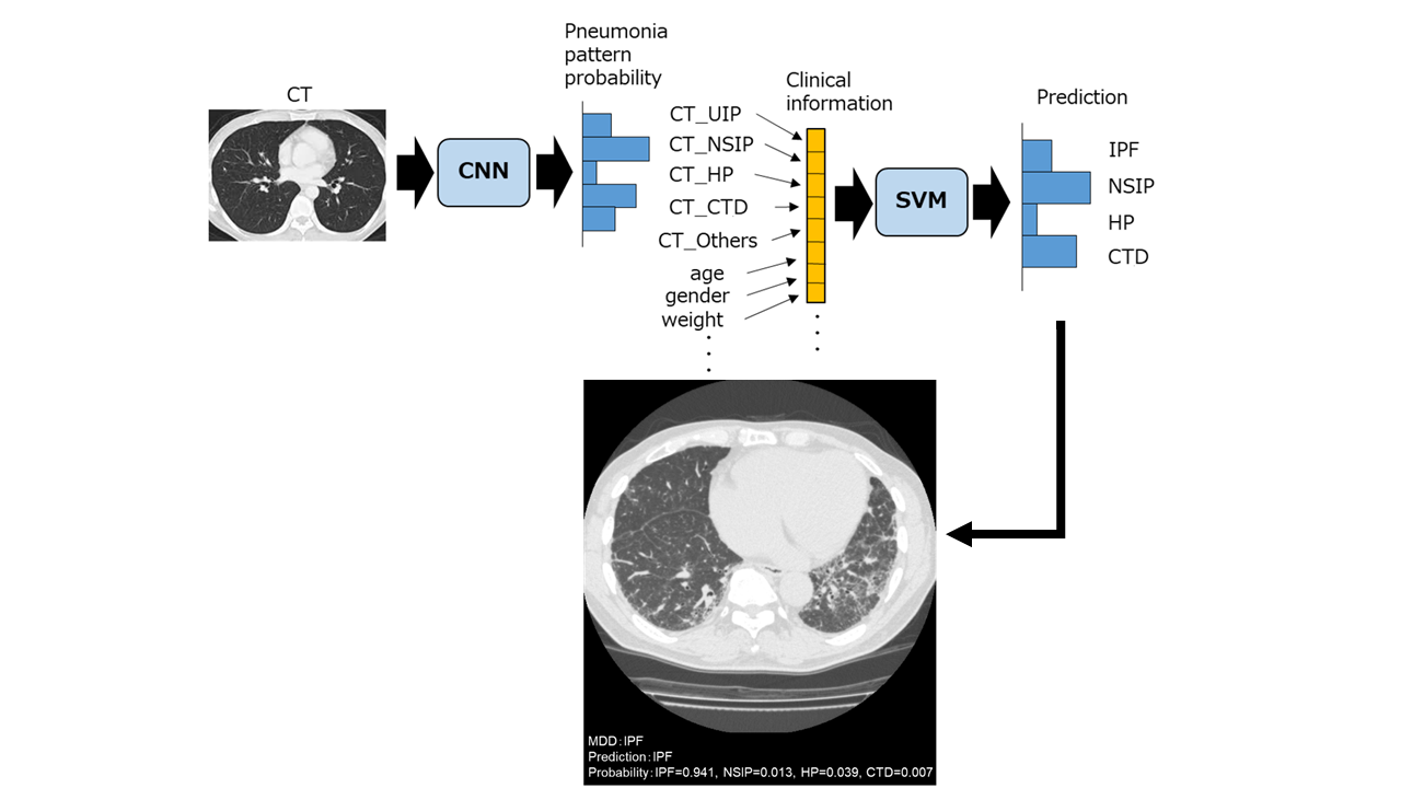
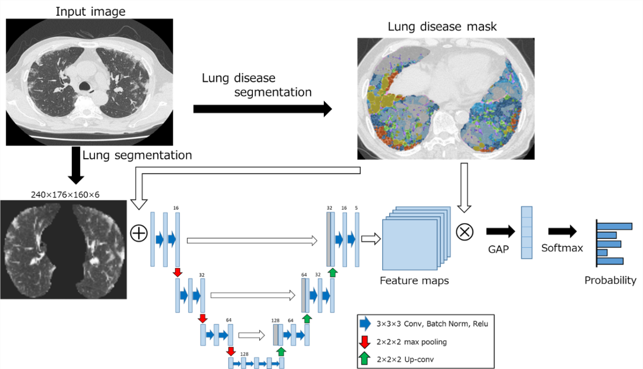
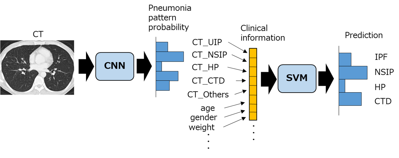
・First stage: pneumonia pattern classification of CT images using a convolutional neural network (CNN) model. (Fig. 1 [1]) This CNN model takes CT images as input and outputs probability scores for five classifications: UIP, NSIP, HP, CTD, and other.
・Second stage: multimodal (clinical, radiological, and pathological) classification using a support vector machine (SVM). (Fig. 2 [1]) When take the output results of the CNN model, clinical information, and pathological diagnosis as input, this SVM model outputs probability scores for four classifications: IPF, NSIP, HP, and CTD.
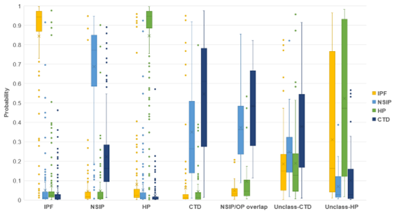
The mean accuracies of the CNN model and SVM were 62.4 % and 85.4 %, respectively. For multimodal classification using SVM, the overall accuracy was very high, especially with sensitivities for idiopathic pulmonary fibrosis and hypersensitivity pneumonitis exceeding 90 %. When pneumonia patterns from CT images, pathological results, or clinical information were not used, the SVM accuracy was 84.3 %, 70.3 % and 79.8 %, respectively, suggesting that pathological results contributed most to MDD diagnosis. The classification results for unclassifiable interstitial pneumonia are for NSIP/OP overlap, it was most commonly classified as CTD, followed by NSIP. Interestingly, unclassifiable interstitial pneumonia tended to be classified by the SVM in line with the most likely diagnosis: “unclass-CTD” to CTD and “unclass-HP” to HP. (Fig.3 [1])
The algorithm based on multimodal information can assist in diagnosing interstitial lung disease and is suitable for ontology diagnosis.
[1] Baba T, Goto T, Kitamura Y, Iwasawa T, Okudela K, Takemura T, et al. (2025) Artificial intelligence for diagnosis in interstitial lung disease and digital ontology for unclassified interstitial lung disease. Respiratory Investigation 63(6): 1179-1186
DOI: https://doi.org/10.1016/j.resinv.2025.09.007
CAUTION:This is Fujifilm Global Website. Fujifilm makes no representation that products on this website are commercially available in all countries. Approved uses of products vary by country and region. Specifications and appearance of products are subject to change without notice.