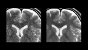

Iterative processing utilizing AI technology to enhance visibility for low dose CT images
The technology controls image quality based on a statistical model, an object model and a physical model using iterative processing.
This technology provides a high visibility image even at a low radiation dose , by maintaining the original texture of the image at a high denoise condition.
It brings both “high visibility at low dose” and “original texture at high noise reduction”
This technology provides a high visibility image even at a low radiation dose , by maintaining the original texture of the image at a high denoise condition.
It brings both “high visibility at low dose” and “original texture at high noise reduction”
Image EnhancementCTITRadiologyCardiologyRespiratoryOrthopedics
Denoising technique utilizing AI technology
We are developing the AI image reconstruction technology that removes noise components of image by preventing the structure and the contrast from degrading by a high speed or a high resolution MR scanning. As a result, the technology shortens the scan time with keeping the image quality, or improves the visibility of the image by improving the image quality.
Image EnhancementMRITRadiologyCardiologyGastroenterologyOrthopedics
Super resolution processing
This flitering technology with AI suppresses aliasing noises which occur in extended images.
Image EnhancementITRadiologyGastroenterology
Noise reduction technology with AI
This technology uses AI to distinguish between echo signals and noises, and extracts the signals necessary for diagnosis. This enables ultrasound systems to provide high-quality images even in difficult cases.
Image EnhancementUSRadiologyCardiologyGastroenterologyOrthopedics
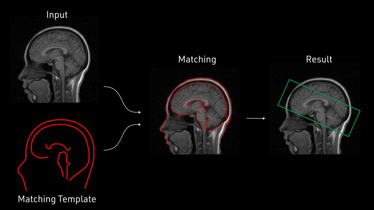
Virtual thin slice generation technology
The technology virtually generates thin slices from thick slices. It can be applied to the whole body, useful for utilizing past data. It brings high visibility in the VR display and reconstructed sagittal/coronal images.
Image EnhancementCTITRadiologyCardiologyRespiratoryGastroenterologyOrthopedics
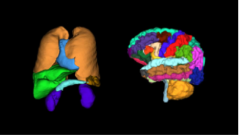
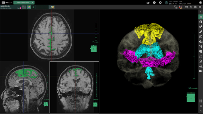
Brain segmentation
AI technology to segment and quantify the volume of each brain regions.
This technology can be used for pre-surgery simulation or calculation of the atrophy rate for each region between past and current exams.
This technology can be used for pre-surgery simulation or calculation of the atrophy rate for each region between past and current exams.
Anatomy SegmentationMRITRadiologyNeurology
Bone temporal subtraction
This technology visualizes the bone density temporal difference by performing image registration between the past and current image of the same patient.
The increase and decrease in CT value will be highlighted.
The increase and decrease in CT value will be highlighted.
Anatomy SegmentationCTITRadiologyOrthopedics
Bone Mineral Density
This technology which developed by deep learning will automatically detect the lumbar spine and femur bone that enhanced by energy subtraction, and calculate the bone mineral density.
Anatomy SegmentationDRRadiology
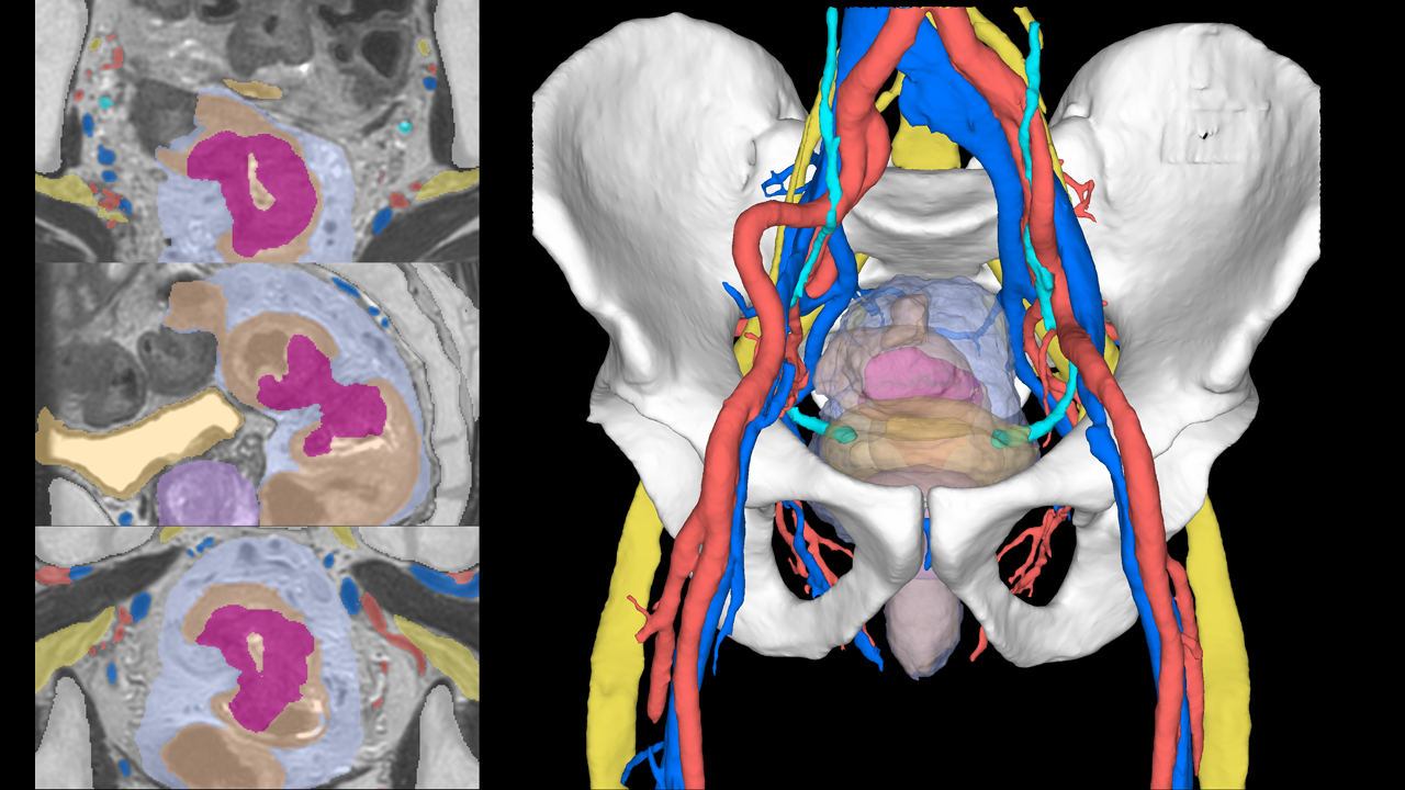
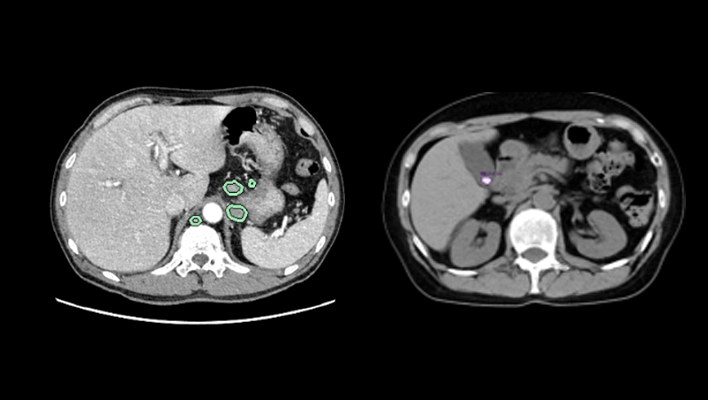
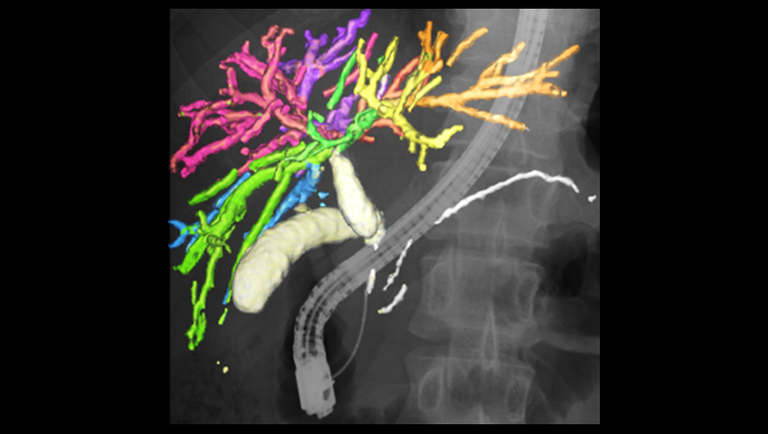
Pancreas analysis
Automatically extracts the pancreas, each surrounding organ area, and surrounding vessels from contrast-enhanced CT images and analyzes them in 3D.
The pancreatic duct diameter, residual pancreatic volume, measurement of the cross-sectional area, and resection simulation can be performed to support pancreatic surgery.
The pancreatic duct diameter, residual pancreatic volume, measurement of the cross-sectional area, and resection simulation can be performed to support pancreatic surgery.
Anatomy SegmentationCTITRadiologyGastroenterology
Kidney segmentation
AI technology to extract each kidney and surrounding vessels from CT or MRI images assists volume calculation of renal and compare current and past results.
This technology can also precisely extract cystic kidneys, a genetic disease that is often difficult to extract due to the diversity of shapes, and assist diagnosis and treatment.
This technology can also precisely extract cystic kidneys, a genetic disease that is often difficult to extract due to the diversity of shapes, and assist diagnosis and treatment.
Anatomy SegmentationCTMRITRadiology
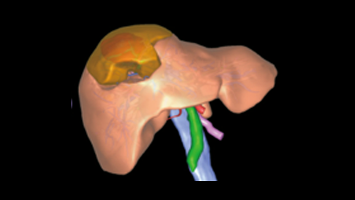
Anatomic liver segments labelling
AI technology to automatically extract liver vessels and then labels liver according to the anatomic liver segments (Couinaud segments) based on the vessel running.
This can be used for Interventional Radiology planning for liver.
This can be used for Interventional Radiology planning for liver.
Anatomy SegmentationCTITRadiologyGastroenterology
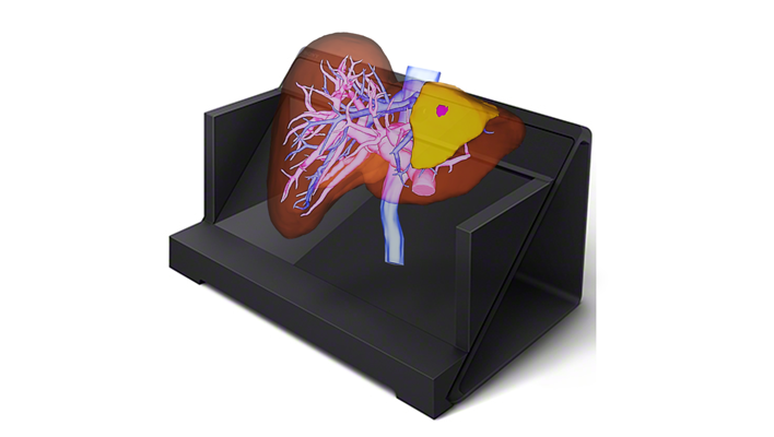
Liver analysis
Technology that automatically extracts the liver parenchyma and surrounding vessels from CT images and analyzes them in 3D.
This technology can assist preoperative simulation by planning resection area based on the calculation of the dominant vessels.
This technology can assist preoperative simulation by planning resection area based on the calculation of the dominant vessels.
Anatomy SegmentationCTITRadiologyGastroenterology
Lung analysis
AI technology to automatically extract lung fields, 5 lobes, and bronchus, surrounding vessels from CT images.
From each extraction result, the percentage of low attenuation area is calculated.
These are expected to contribute the diagnosis of COPD.
From each extraction result, the percentage of low attenuation area is calculated.
These are expected to contribute the diagnosis of COPD.
Anatomy SegmentationCTITRadiologyRespiratory
Knee joint analysis
This technology automatically extracts the femur, tibia, patella, cartilage, and meniscus from MRI images.
By measuring the thickness of the cartilage and the distance between the meniscus and tibia, it is possible to evaluate the condition of the knee joint without invasion.
By measuring the thickness of the cartilage and the distance between the meniscus and tibia, it is possible to evaluate the condition of the knee joint without invasion.
Anatomy SegmentationMRITRadiologyOrthopedics
Vertebrae and ribs labelling
Technology to recognize each vertebral body individually and label the vertebrae and ribs. This reduces the burden on the physician of visually counting bone positions. It is also used for visualization of fractures and bone metastasis by subtraction of rigid body alignment of vertebral bodies.
Anatomy SegmentationCTITRadiologyOrthopedics
Lung labelling
AI technology to subdivide lung into 10 segments in right lung, 8 segments in left lung from CT images. It can be used for confirming the position of chest nodules detected by a doctor.
Anatomy SegmentationCTITRadiologyRespiratory
Mediastinum and axillary lymph node segmentation
AI technology to segment relatively large mediastinum, axillary lymph node from CT images. This technology can be used for assessment of lymph node enlargement.
Anatomy SegmentationCTITRadiology
Rib fracture CAD
AI technology to detect a suspcious rib fracture from CT images. This technology will assist prevention of overlooking of subtle rib fracture.
Detection/DiagnosisCTITRadiology
COVID CAD
AI technology to identify suspicious region with COVID-19 related findings from CT images. This technology will help doctors diagnose efficiently.
Detection/DiagnosisCTITRadiologyRespiratory
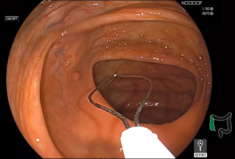
Detection assist technology for colonic polyps
This technology assists real time detection and characterization of colonic polyps from colonoscopy images with AI software.
Detection/DiagnosisESGastroenterology
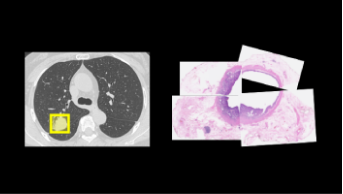
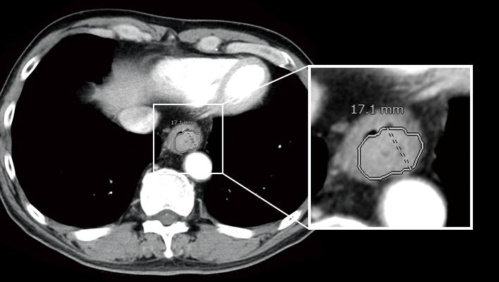
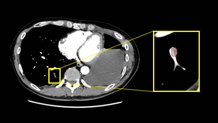
Lung nodule CAD
AI technology to detect and quantify suspicious lesion from CT Images.
This will assit prevention of overlooking of nodule and generation of language of findings for radiology report.
This will assit prevention of overlooking of nodule and generation of language of findings for radiology report.
Detection/DiagnosisCTITRadiologyRespiratory
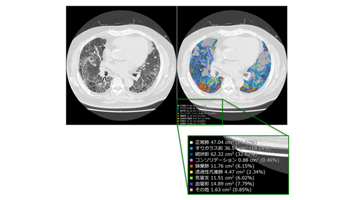
Interstitial lung disease classification
This technology identifies various findings of interstitial pneumonia that appear on CT images, such as consolidation, reticular pattern, ground glass opacity and honeycomb, and calculates their distribution and volume.
This will assist in the diagnosis of the severity and therapeutic efficacy of interstitial pneumonia, which are conventionally performed qualitatively, by providing a quantitative value for assessment.
This will assist in the diagnosis of the severity and therapeutic efficacy of interstitial pneumonia, which are conventionally performed qualitatively, by providing a quantitative value for assessment.
Detection/DiagnosisCTITRadiologyRespiratory
AI-CAD in modality
The technology which prepares environment on the modality to exapnd the opportuities of AI-CAD applications and improve workflow not only in hospitals, but also at point of care.
Detection/DiagnosisDROthers
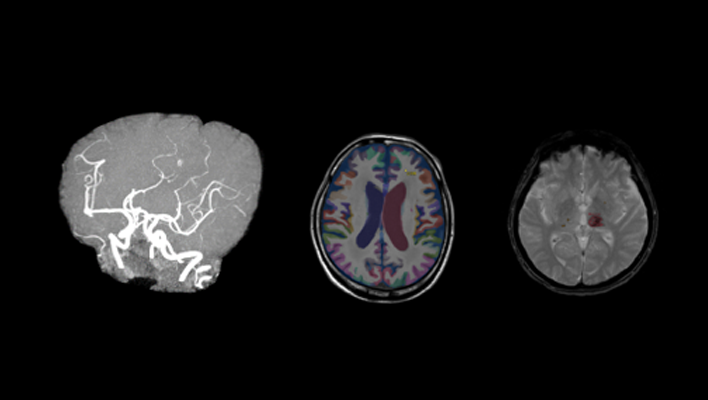
Highlighting areas of higher or lower signals by comparing left/right head CT images
This technology extracts high-signal and low-signal areas in head CT images by comparing the left and right sides of the brain region. Generally, high-signal and low-signal areas are used to evaluate the state of hemorrhage and ischemia in the brain for stroke diagnosis. This will assist the diagnosis of head CT imaging.
Detection/DiagnosisCTITRadiologyNeurology
Highlighting areas of higher or lower absorption than the surrounding tissue
The technology to extract and highlight areas of higher or lower absorption than the surrounding tissue. For example, high/low absorption in the liver, high absorption in the thoracic cavity, and low absorption in the kidney may be informative in determining the findings of each part. In the future, we aim to highlight both contrast-enhanced and non-contrast-enhanced images.
Detection/DiagnosisCTITRadiology
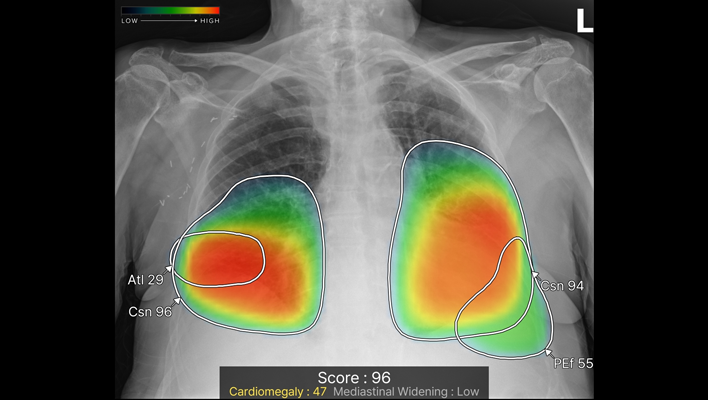
Chest X-ray CAD
The technology detects ten types of imaging findings: nodule, consolidation, pneumothorax, atelectasis, calcification, scar, pleural effusion, pneumoperitoneum, cardiomegaly and mediastinal widening from chest X-ray images. It is expected to contribute to preventing oversights in various chest X-ray examinations, such as health checkups and routine medical examinations.
Detection/DiagnosisITRadiologyRespiratory
Real-time Screening Assist
This technology highlights areas in the B-mode image where luminance characteristics are different from the surroundings
Detection/DiagnosisUSRadiology
Improvement of workflow with AI
This technology extracts the target area from the image.
Positioning during imaging is performed automatically.
Unnecessary tissue is automatically removed before creating a 3D image for diagnosis.
It is a technology that supports the automation of inspections.
Positioning during imaging is performed automatically.
Unnecessary tissue is automatically removed before creating a 3D image for diagnosis.
It is a technology that supports the automation of inspections.
Workflow SupportMRITRadiology
Automatic view recognition,Automatic measurement
The technology enables automatic recognition of left ventricle, left atrium and right atrium wall in addition to view recognition of the heart, and automatic measurements realize autonomous measurements of ejection fraction and volumes.
The measurement function are used very often in echocardiography exams. This AI technology assist to reduce inter- and intra-observer variability so that improve reproducibility and efficiency of exams.
The measurement function are used very often in echocardiography exams. This AI technology assist to reduce inter- and intra-observer variability so that improve reproducibility and efficiency of exams.
Workflow SupportUSCardiology
Blood vessel detection technology
This technology assists real time detection and distinction between an artery and a vein, as well as measurement of diameter and depth of the vessel from ultrasound images. This will streamline Point Of Care UltraSound exams.
Workflow SupportUSOthers
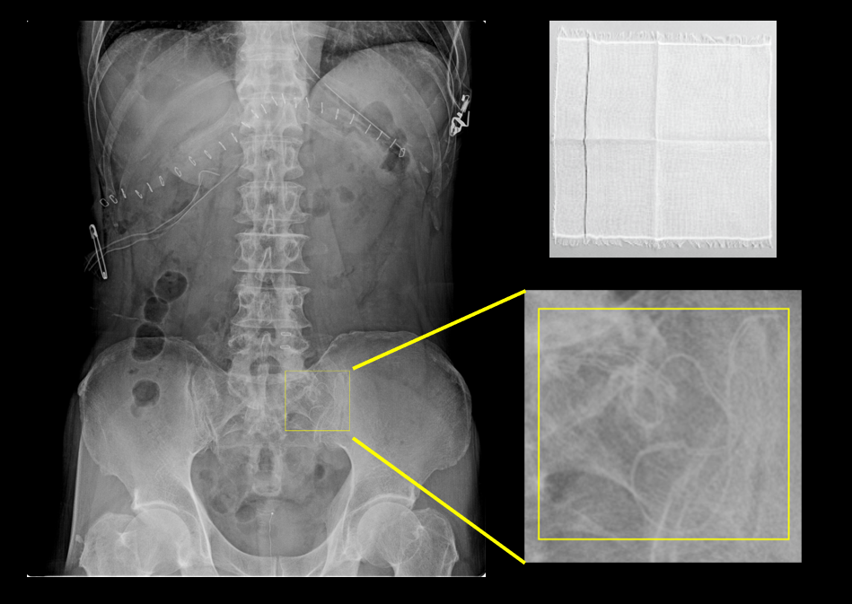
Retained foreign object detection
This AI technology detects foreign objects in X-ray images. This technology can be applied to the detection of surgical objects such as gauze and reduce the burden on doctors to check the surgical objects held at each surgical procedure.
Workflow SupportDROthers
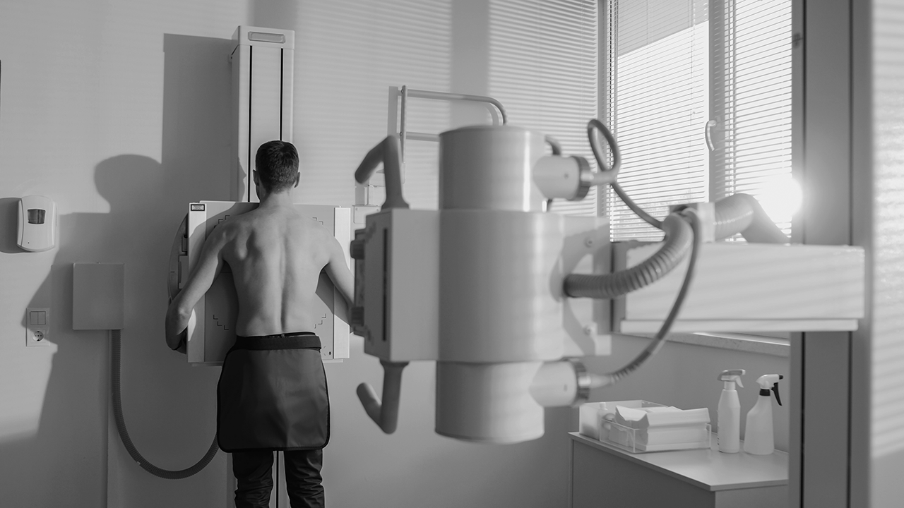
Positioning assist
This technology is to assist positioning before exposure by images from camera which implemented on the X-ray modality and confirming the more optimal positioin with AI software .
Workflow SupportDRRadiology
Exposure condition navigation
The sensor integrated in the X-ray system will recognize three-dimensional information from the patient's body.
It will then automatically suggest the optimized exposure conditions and adapt proper image processing.
It will then automatically suggest the optimized exposure conditions and adapt proper image processing.
Workflow SupportDRRadiology
Prediction of clinical outcomes using inpatient data with pneumonia
We developed AI algorithms to predict clinically important outcomes using inpatient data with pneumonia aggregated throught our integrated medical care support platform system. This technology will assist preparing personalized medical documents and optimizing medical resources
Workflow SupportITOthers
Lung nodule characterization analysis
The technology supports populating radiology report of lung nodules based on the observation and analysis results of the properties. It enables to analyze multiple properties such as shape, absorption, presence of calcification, etc., and provide multiple finding sentence candidates using natural language processing technology.
Workflow SupportCTITRadiology
Liver characterization analysis
The technology analyzes the properties of areas of interest in the liver and assists doctors in writing radiology report. It is possible to analyze not only the location and size, but also the appearance/disappearance of enhancement based on multi-phase images of contrast-enhanced CT. In addition, similar to lung nodule characterization analysis, it is also possible to present multiple finding sentence candidates using natural language processing technology.
Workflow SupportCTITRadiology
Quantification of high absorption ROI in lung field
The technology estimates high-value threshold of the region of interest, and quantify high intensity area in lung field. For example, it is expected to offer information for quantitative analysis of partially solid nodules.
Detection/DiagnosisCTITRadiologyRespiratory
Automatic measurement of EvansIndex, corpus callosum angle, and MidlineShift
This technology automatically measures EvansIndex, corpus callosum angle, and MidlineShift from head CT. EvansIndex and corpus callosum angle are expected to support the evaluation of hydrocephalus and MidlineShift to evaluate head trauma.
Detection/DiagnosisCTITRadiologyNeurology
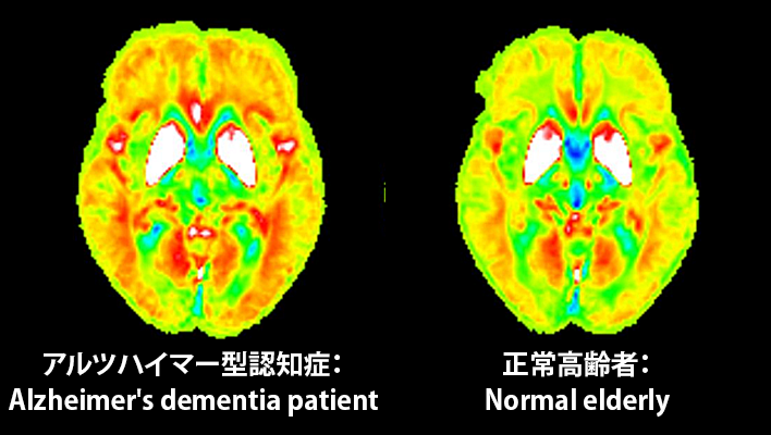
Early diagnosis of dementia
The technology under development quantitative detection of slight brain volume loss and iron deposition in the early stages of dementia using hybrid analysis of QSM(Quantitative Susceptibility Mapping)and VBM(Voxel Based Morphometry)in MRI. This will assist the diagnosis of dementia.
Detection/DiagnosisMRITRadiologyNeurology
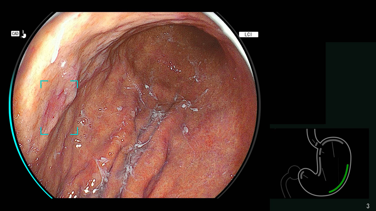
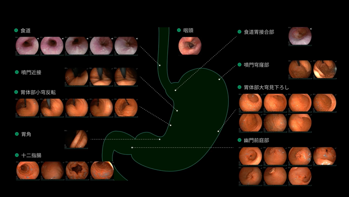
Detection technology for gastric neoplastic lesions and suspected esophageal squamous cell carcinoma
The technology utilizes AI to analyze upper gastrointestinal endoscopic images, recognizing areas suspected of being gastric neoplastic lesions or esophageal squamous cell carcinoma and detecting them in real time.
Detection/DiagnosisESGastroenterology
Endoscopy report creation support technology
In lower gastrointestinal endoscopy, AI technology is used to automatically recognize the insertion and removal of the scope from the body, as well as the insertion of surgical instruments, through software.
This is expected to support the recording of examination times and information on specimen collection (e.g., polyps), thereby reducing the burden on physicians in preparing reports.
This is expected to support the recording of examination times and information on specimen collection (e.g., polyps), thereby reducing the burden on physicians in preparing reports.
Workflow SupportESGastroenterology
AI technology for abdominal CT
Technology that displays areas of high and low absorption in the liver, kidneys, and spleen compared to surrounding tissues.
Anatomy SegmentationCTITRadiology
Pelvic segmentation technology
The technology for segmenting the rectum and surrounding organs from MRI images, which is expected to be useful for surgical simulation in the lower digestive tract area.
Anatomy SegmentationMRITRadiologyGastroenterology
Pancreatic cancer detection technology
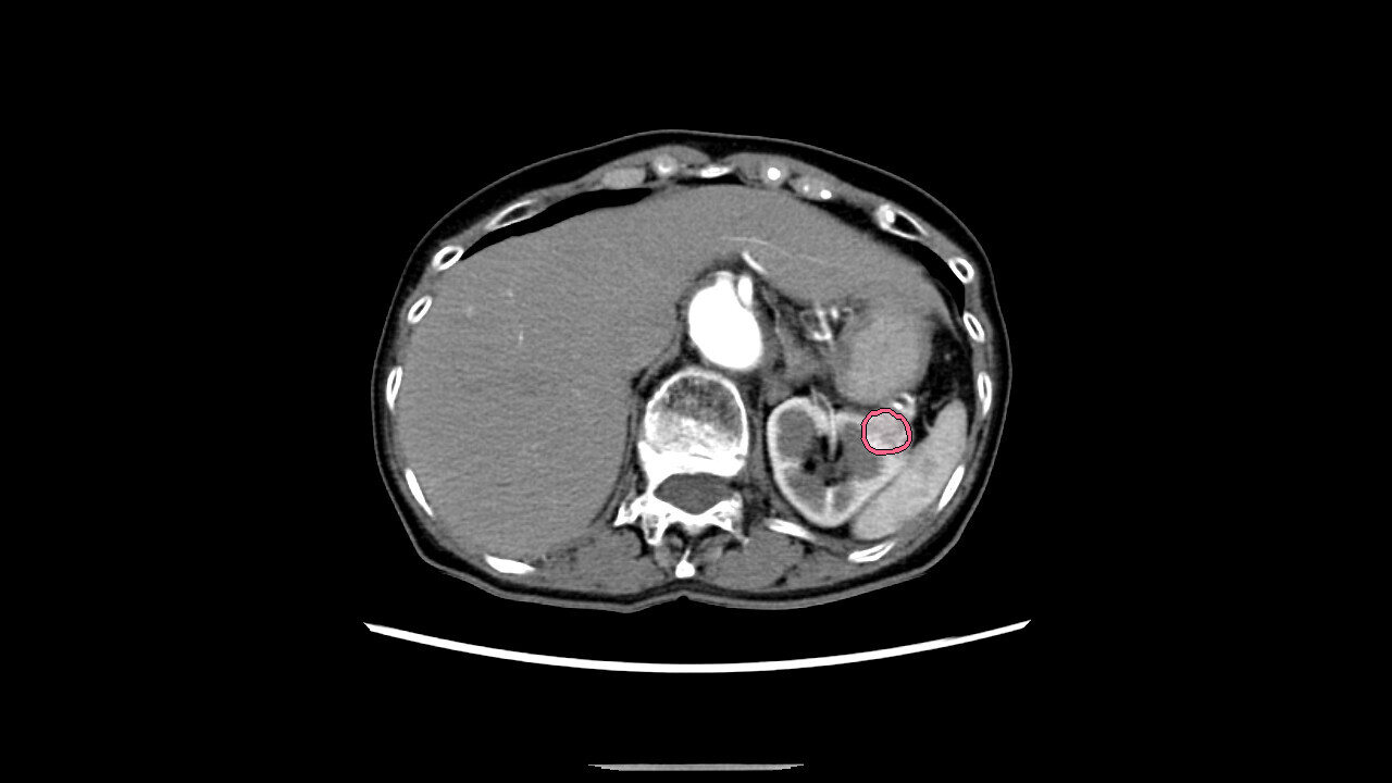
The technology that utilizes AI to help detect findings suggestive of pancreatic cancer from abdominal CT images.
Detection/DiagnosisITRadiologyGastroenterology
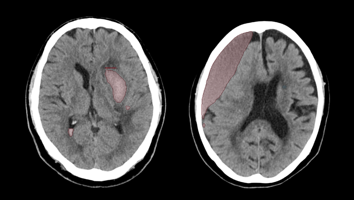
CT technology for diagnosing subarachnoid hemorrhage
We have developed AI technology to identify areas suspected of cerebral hemorrhage or cerebral infarction in CT images of the head.
This technology is expected to aid in the diagnosis of stroke by helping to evaluate bleeding and ischemia in the brain.
This technology is expected to aid in the diagnosis of stroke by helping to evaluate bleeding and ischemia in the brain.
Detection/DiagnosisCTITRadiologyNeurology
Cerebrospinal fluid analysis technology in MRI images
We have developed AI technology to extract each region of the cerebrospinal fluid cavity from MRI images.
The aim is to improve diagnostic accuracy for Hakim's disease (iNPH), a form of dementia that is important to detect early for treatment.
This technology is expected to improve the objectivity and accuracy of diagnosis by enabling AI to efficiently analyze regions associated with key findings (DESH) and support differentiation from brain atrophy.
Anatomy SegmentationMRITRadiologyNeurology
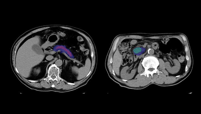
Pulmonary artery absorption enhancement technique
The technology that displays areas of low absorption in the pulmonary artery compared to surrounding tissues.
This is expected to aid in the diagnosis of pulmonary embolism.
This is expected to aid in the diagnosis of pulmonary embolism.
Anatomy SegmentationCTITRadiologyCardiologyRespiratory
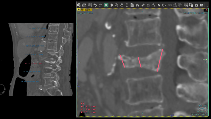
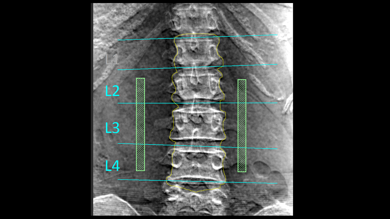
Automatic vertebral body height measurement technology
The technology that automatically measures the height of each vertebral body and displays the results classified according to user-defined thresholds.
This technology is expected to aid in the diagnosis of vertebral fractures.
This technology is expected to aid in the diagnosis of vertebral fractures.
Anatomy SegmentationITRadiologyOrthopedics
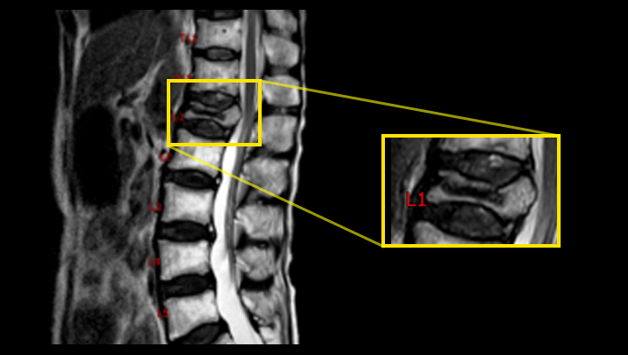
MRI labeling technology
The technique for labeling vertebral numbers on MRI images.
Anatomy SegmentationMRITRadiologyOrthopedics
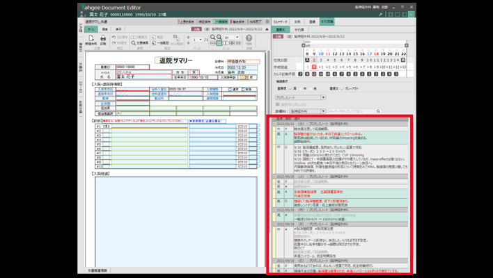
Medical record summary support technology
When creating documents such as discharge summaries, we developed a technology that extracts necessary text from medical records to assist.
Workflow SupportITOthers
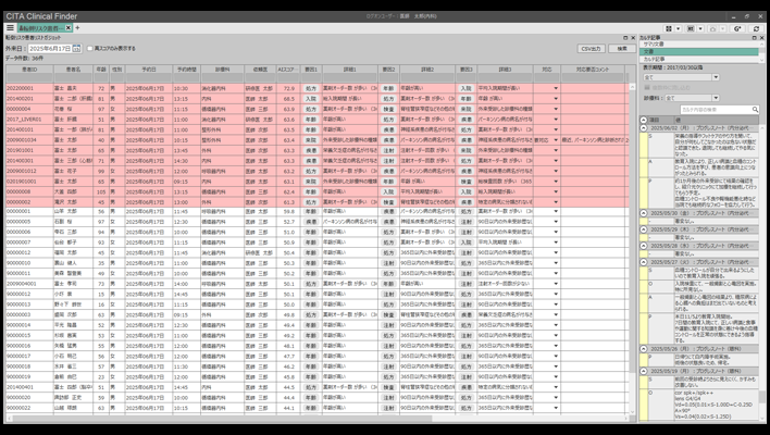
AI technology for predicting fall risk in outpatients
Based on clinical data accumulated in “CITA Clinical Finder,”
we have developed technology that predicts the risk of falls among outpatients, provides risk scores, and identifies factors contributing to AI predictions.
we have developed technology that predicts the risk of falls among outpatients, provides risk scores, and identifies factors contributing to AI predictions.
Workflow SupportITOthers
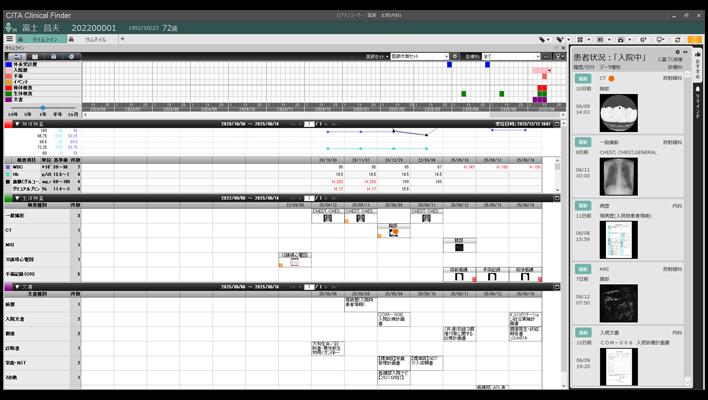
AI technology for medical data reference support
We have developed technology that predicts and lists medical data that is likely to be viewed based on patient information accumulated in “CITA Clinical Finder” and the viewing status of users, thereby supporting the reference of medical data.
Workflow SupportITOthers
AI technology for abdominal CT
The technology that highlights areas of high/low absorption in the adrenal glands, pancreas, gallbladder, and lymph nodes compared to surrounding tissues.
Anatomy SegmentationCTITRadiologyGastroenterology
AI technology for head MRI
High signal/low signal region extraction technology, brain region labeling technology, brain extraction technology.
Anatomy SegmentationMRITRadiologyNeurology
Liver deformation technology
Observe the liver and surrounding organs while deforming them, enabling estimation of the position of blood vessels during surgical detachment.
Anatomy SegmentationITRadiologyGastroenterology
Naked-eye stereoscopic technology
Display 3D images created in the target application on a spatial reproduction display, and operate them in synchronization with our 3D system.
Anatomy SegmentationITRadiologyCardiologyRespiratoryGastroenterologyOrthopedics
AI technology for the gallbladder and pancreas
Displays the complex structure of the gallbladder and pancreas in three dimensions, supporting preoperative simulation of organ relationships and other factors.
Anatomy SegmentationITRadiologyGastroenterology
Endoscopy report creation support technology
In upper gastrointestinal endoscopy, AI technology is used to automatically recognize the location of images taken by doctors.
By extracting appropriate images for each location and automatically recording them in a report, it is expected that the burden of report creation for doctors will be reduced.
By extracting appropriate images for each location and automatically recording them in a report, it is expected that the burden of report creation for doctors will be reduced.
Workflow SupportESGastroenterology
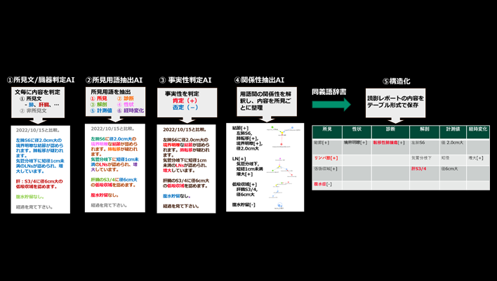
Natural language processing technology “AI for structuring medical reports”
Fujifilm has developed “Reading Report Structuring AI,” a unique natural language processing technology that structures reading reports created by radiologists for medical images captured by CT, MRI, and other imaging devices.
By structuring reading reports written based on the specialized knowledge of radiologists, it is possible to efficiently database and utilize reading reports that include medical jargon and expressions unique to physicians.
By structuring reading reports written based on the specialized knowledge of radiologists, it is possible to efficiently database and utilize reading reports that include medical jargon and expressions unique to physicians.
Workflow SupportITOthers
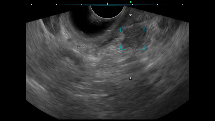
Ultrasound endoscopic diagnostic support technology for the pancreatic region
We have developed ultrasound endoscopy diagnostic support software that detects areas suspected of pancreatic solid lesions in real time during ultrasound endoscopy examinations, thereby supporting the early detection of pancreatic cancer.
By analyzing ultrasound endoscopy images, the software detects areas where the pancreas is presumed to exist and areas suspected of pancreatic solid lesions in real time, and displays the results on the monitor's ultrasound endoscopy image.
By alerting the operator, the software assists in detecting pancreatic solid lesions.
Detection/DiagnosisESRadiologyGastroenterology
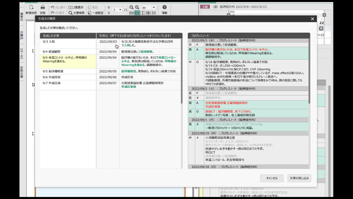
Summary creation support technology
The technology utilizes a large-scale language model to automatically generate and present drafts of "summaries," which are documents that comprehensively describe patient information such as test results and treatment history.
Workflow SupportITOthers
AI technology for classifying the characteristics of areas suspected of interstitial lung disease and calculating their size
This AI technology identifies the anatomical structures of the lungs from chest CT images, automatically classifies characteristics based on imaging feature patterns, and simultaneously calculates their size and proportion. Additionally, to enable detailed review of the distribution of abnormalities, the lung fields are segmented by lobes, with the size and proportion of abnormalities in each region displayed.
Detection/DiagnosisITRespiratory
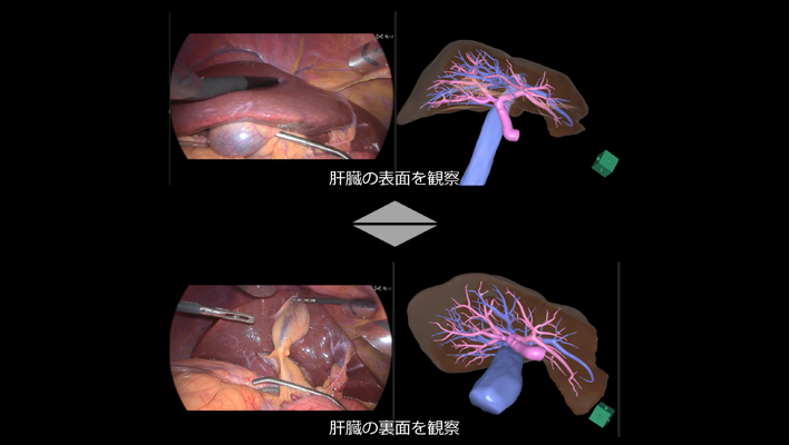
AI technology linked with 3D liver imaging
An AI technology that automatically rotates the preoperatively created 3D liver image to the same orientation as the liver in endoscopic video, allowing simultaneous reference on the same monitor, thereby supporting the understanding of internal liver structures such as blood vessels and tumor locations that are difficult to observe from the liver surface.
Workflow SupportITGastroenterology
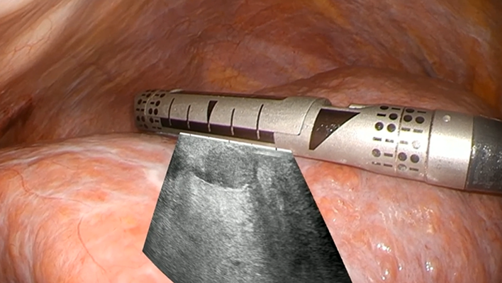
AI technology for ultrasound image overlay
An AI technology that analyzes the position and orientation of the ultrasound probe within endoscopic images during intraoperative ultrasound examinations performed in laparoscopic or robot-assisted surgeries, and overlays the ultrasound images onto the endoscopic video to support intuitive understanding of the ultrasound images.
Workflow SupportITGastroenterology