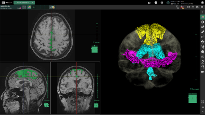
Brain segmentation
AI technology to segment and quantify the volume of each brain regions.
This technology can be used for pre-surgery simulation or calculation of the atrophy rate for each region between past and current exams.
This technology can be used for pre-surgery simulation or calculation of the atrophy rate for each region between past and current exams.
Anatomy SegmentationMRITRadiologyNeurology
Bone temporal subtraction
This technology visualizes the bone density temporal difference by performing image registration between the past and current image of the same patient.
The increase and decrease in CT value will be highlighted.
The increase and decrease in CT value will be highlighted.
Anatomy SegmentationCTITRadiologyOrthopedics
Bone Mineral Density
This technology which developed by deep learning will automatically detect the lumbar spine and femur bone that enhanced by energy subtraction, and calculate the bone mineral density.
Anatomy SegmentationDRRadiology
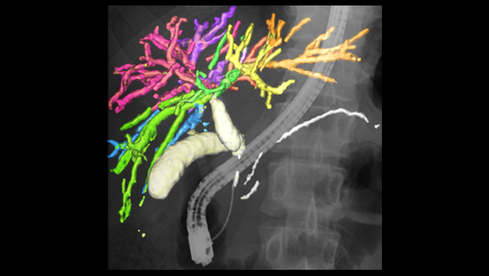
Pancreas analysis
Automatically extracts the pancreas, each surrounding organ area, and surrounding vessels from contrast-enhanced CT images and analyzes them in 3D.
The pancreatic duct diameter, residual pancreatic volume, measurement of the cross-sectional area, and resection simulation can be performed to support pancreatic surgery.
The pancreatic duct diameter, residual pancreatic volume, measurement of the cross-sectional area, and resection simulation can be performed to support pancreatic surgery.
Anatomy SegmentationCTITRadiologyGastroenterology
Kidney segmentation
AI technology to extract each kidney and surrounding vessels from CT or MRI images assists volume calculation of renal and compare current and past results.
This technology can also precisely extract cystic kidneys, a genetic disease that is often difficult to extract due to the diversity of shapes, and assist diagnosis and treatment.
This technology can also precisely extract cystic kidneys, a genetic disease that is often difficult to extract due to the diversity of shapes, and assist diagnosis and treatment.
Anatomy SegmentationCTMRITRadiology
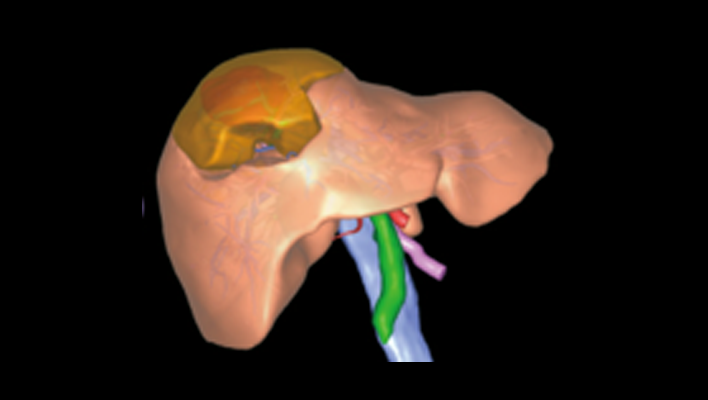
Anatomic liver segments labelling
AI technology to automatically extract liver vessels and then labels liver according to the anatomic liver segments (Couinaud segments) based on the vessel running.
This can be used for Interventional Radiology planning for liver.
This can be used for Interventional Radiology planning for liver.
Anatomy SegmentationCTITRadiologyGastroenterology
Liver analysis
Technology that automatically extracts the liver parenchyma and surrounding vessels from CT images and analyzes them in 3D.
This technology can assist preoperative simulation by planning resection area based on the calculation of the dominant vessels.
This technology can assist preoperative simulation by planning resection area based on the calculation of the dominant vessels.
Anatomy SegmentationCTITRadiologyGastroenterology
Lung analysis
AI technology to automatically extract lung fields, 5 lobes, and bronchus, surrounding vessels from CT images.
From each extraction result, the percentage of low attenuation area is calculated.
These are expected to contribute the diagnosis of COPD.
From each extraction result, the percentage of low attenuation area is calculated.
These are expected to contribute the diagnosis of COPD.
Anatomy SegmentationCTITRadiologyRespiratory
Knee joint analysis
This technology automatically extracts the femur, tibia, patella, cartilage, and meniscus from MRI images.
By measuring the thickness of the cartilage and the distance between the meniscus and tibia, it is possible to evaluate the condition of the knee joint without invasion.
By measuring the thickness of the cartilage and the distance between the meniscus and tibia, it is possible to evaluate the condition of the knee joint without invasion.
Anatomy SegmentationMRITRadiologyOrthopedics
Vertebrae and ribs labelling
Technology to recognize each vertebral body individually and label the vertebrae and ribs. This reduces the burden on the physician of visually counting bone positions. It is also used for visualization of fractures and bone metastasis by subtraction of rigid body alignment of vertebral bodies.
Anatomy SegmentationCTITRadiologyOrthopedics
Lung labelling
AI technology to subdivide lung into 10 segments in right lung, 8 segments in left lung from CT images. It can be used for confirming the position of chest nodules detected by a doctor.
Anatomy SegmentationCTITRadiologyRespiratory
Mediastinum and axillary lymph node segmentation
AI technology to segment relatively large mediastinum, axillary lymph node from CT images. This technology can be used for assessment of lymph node enlargement.
Anatomy SegmentationCTITRadiology
AI technology for abdominal CT
Technology that displays areas of high and low absorption in the liver, kidneys, and spleen compared to surrounding tissues.
Anatomy SegmentationCTITRadiology
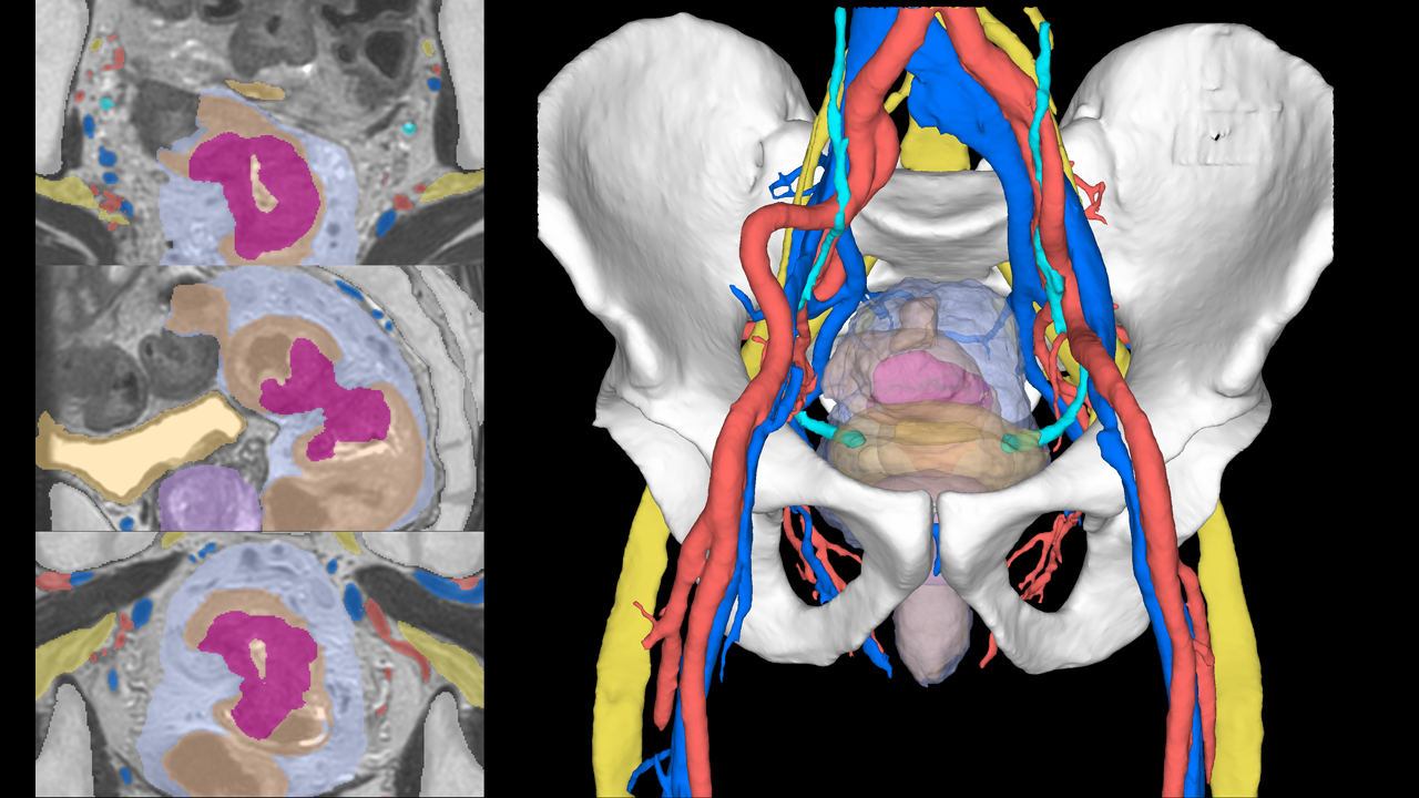
Pelvic segmentation technology
The technology for segmenting the rectum and surrounding organs from MRI images, which is expected to be useful for surgical simulation in the lower digestive tract area.
Anatomy SegmentationMRITRadiologyGastroenterology
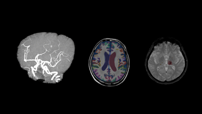
Cerebrospinal fluid analysis technology in MRI images
We have developed AI technology to extract each region of the cerebrospinal fluid cavity from MRI images.
The aim is to improve diagnostic accuracy for Hakim's disease (iNPH), a form of dementia that is important to detect early for treatment.
This technology is expected to improve the objectivity and accuracy of diagnosis by enabling AI to efficiently analyze regions associated with key findings (DESH) and support differentiation from brain atrophy.
Anatomy SegmentationMRITRadiologyNeurology
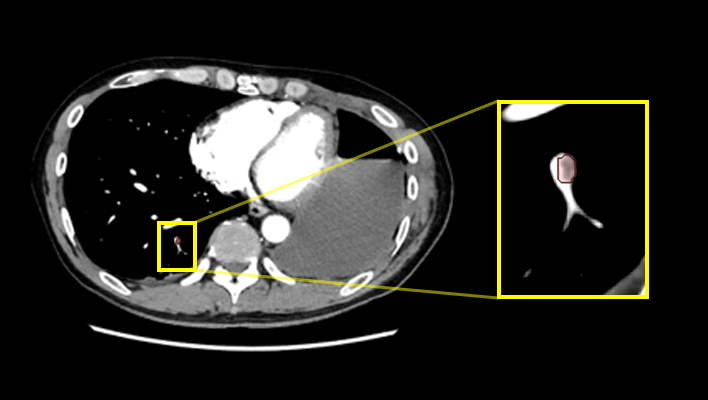
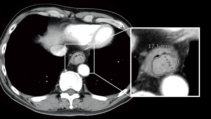
Pulmonary artery absorption enhancement technique
The technology that displays areas of low absorption in the pulmonary artery compared to surrounding tissues.
This is expected to aid in the diagnosis of pulmonary embolism.
This is expected to aid in the diagnosis of pulmonary embolism.
Anatomy SegmentationCTITRadiologyCardiologyRespiratory
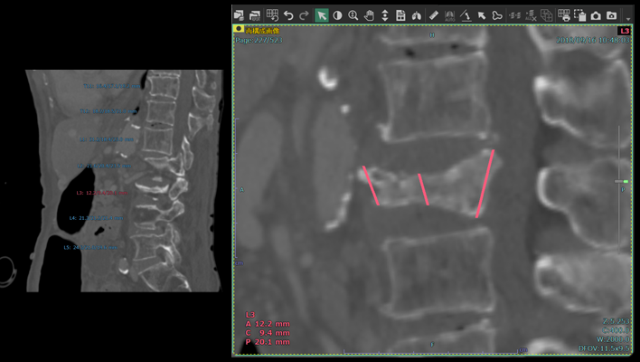
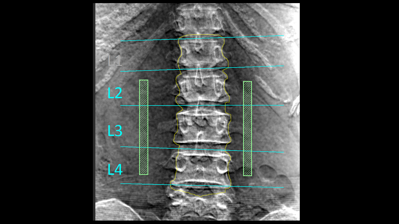
Automatic vertebral body height measurement technology
The technology that automatically measures the height of each vertebral body and displays the results classified according to user-defined thresholds.
This technology is expected to aid in the diagnosis of vertebral fractures.
This technology is expected to aid in the diagnosis of vertebral fractures.
Anatomy SegmentationITRadiologyOrthopedics
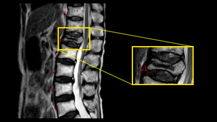
MRI labeling technology
The technique for labeling vertebral numbers on MRI images.
Anatomy SegmentationMRITRadiologyOrthopedics
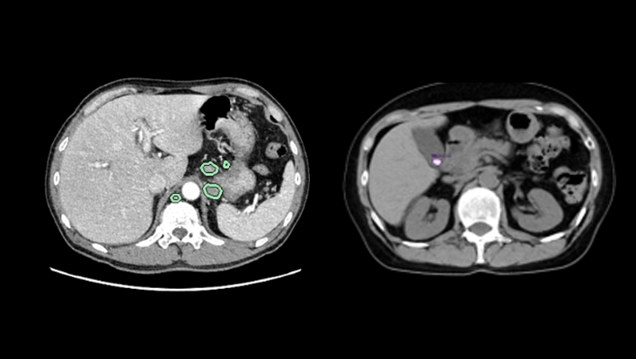
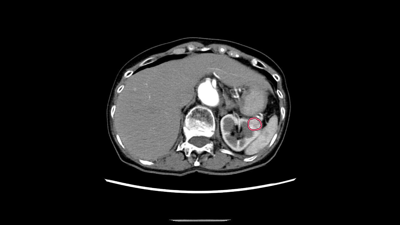
AI technology for abdominal CT
The technology that highlights areas of high/low absorption in the adrenal glands, pancreas, gallbladder, and lymph nodes compared to surrounding tissues.
Anatomy SegmentationCTITRadiologyGastroenterology
AI technology for head MRI
High signal/low signal region extraction technology, brain region labeling technology, brain extraction technology.
Anatomy SegmentationMRITRadiologyNeurology
Liver deformation technology
Observe the liver and surrounding organs while deforming them, enabling estimation of the position of blood vessels during surgical detachment.
Anatomy SegmentationITRadiologyGastroenterology
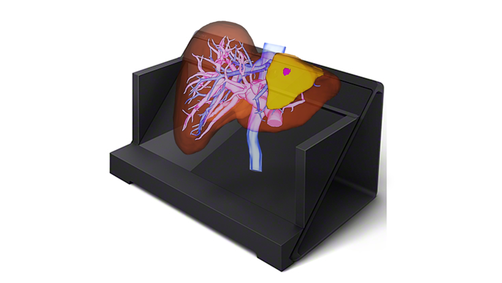
Naked-eye stereoscopic technology
Display 3D images created in the target application on a spatial reproduction display, and operate them in synchronization with our 3D system.
Anatomy SegmentationITRadiologyCardiologyRespiratoryGastroenterologyOrthopedics
AI technology for the gallbladder and pancreas
Displays the complex structure of the gallbladder and pancreas in three dimensions, supporting preoperative simulation of organ relationships and other factors.
Anatomy SegmentationITRadiologyGastroenterology