Iterative processing utilizing AI technology to enhance visibility for low dose CT images
The technology controls image quality based on a statistical model, an object model and a physical model using iterative processing.
This technology provides a high visibility image even at a low radiation dose , by maintaining the original texture of the image at a high denoise condition.
It brings both “high visibility at low dose” and “original texture at high noise reduction”
This technology provides a high visibility image even at a low radiation dose , by maintaining the original texture of the image at a high denoise condition.
It brings both “high visibility at low dose” and “original texture at high noise reduction”
Image EnhancementCTITRadiologyCardiologyRespiratoryOrthopedics
Denoising technique utilizing AI technology
We are developing the AI image reconstruction technology that removes noise components of image by preventing the structure and the contrast from degrading by a high speed or a high resolution MR scanning. As a result, the technology shortens the scan time with keeping the image quality, or improves the visibility of the image by improving the image quality.
Image EnhancementMRITRadiologyCardiologyGastroenterologyOrthopedics
Noise reduction technology with AI
This technology uses AI to distinguish between echo signals and noises, and extracts the signals necessary for diagnosis. This enables ultrasound systems to provide high-quality images even in difficult cases.
Image EnhancementUSRadiologyCardiologyGastroenterologyOrthopedics
Virtual thin slice generation technology
The technology virtually generates thin slices from thick slices. It can be applied to the whole body, useful for utilizing past data. It brings high visibility in the VR display and reconstructed sagittal/coronal images.
Image EnhancementCTITRadiologyCardiologyRespiratoryGastroenterologyOrthopedics
Bone temporal subtraction
This technology visualizes the bone density temporal difference by performing image registration between the past and current image of the same patient.
The increase and decrease in CT value will be highlighted.
The increase and decrease in CT value will be highlighted.
Anatomy SegmentationCTITRadiologyOrthopedics
Knee joint analysis
This technology automatically extracts the femur, tibia, patella, cartilage, and meniscus from MRI images.
By measuring the thickness of the cartilage and the distance between the meniscus and tibia, it is possible to evaluate the condition of the knee joint without invasion.
By measuring the thickness of the cartilage and the distance between the meniscus and tibia, it is possible to evaluate the condition of the knee joint without invasion.
Anatomy SegmentationMRITRadiologyOrthopedics
Vertebrae and ribs labelling
Technology to recognize each vertebral body individually and label the vertebrae and ribs. This reduces the burden on the physician of visually counting bone positions. It is also used for visualization of fractures and bone metastasis by subtraction of rigid body alignment of vertebral bodies.
Anatomy SegmentationCTITRadiologyOrthopedics
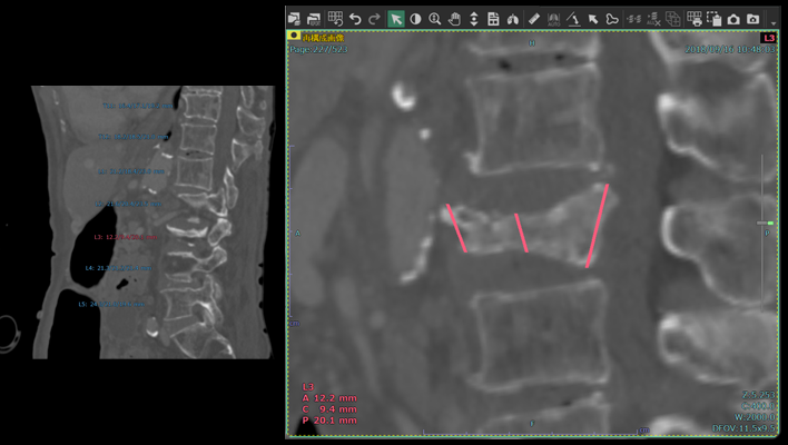
Automatic vertebral body height measurement technology
The technology that automatically measures the height of each vertebral body and displays the results classified according to user-defined thresholds.
This technology is expected to aid in the diagnosis of vertebral fractures.
This technology is expected to aid in the diagnosis of vertebral fractures.
Anatomy SegmentationITRadiologyOrthopedics
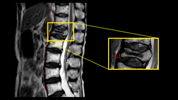
MRI labeling technology
The technique for labeling vertebral numbers on MRI images.
Anatomy SegmentationMRITRadiologyOrthopedics
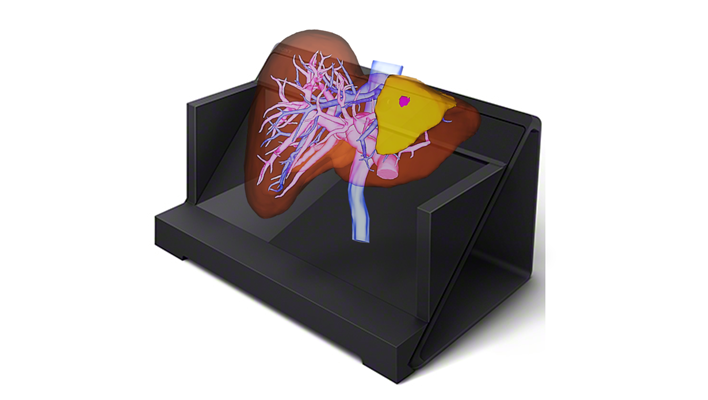
Naked-eye stereoscopic technology
Display 3D images created in the target application on a spatial reproduction display, and operate them in synchronization with our 3D system.
Anatomy SegmentationITRadiologyCardiologyRespiratoryGastroenterologyOrthopedics