Denoising technique utilizing AI technology
We are developing the AI image reconstruction technology that removes noise components of image by preventing the structure and the contrast from degrading by a high speed or a high resolution MR scanning. As a result, the technology shortens the scan time with keeping the image quality, or improves the visibility of the image by improving the image quality.
Image EnhancementMRITRadiologyCardiologyGastroenterologyOrthopedics
Super resolution processing
This flitering technology with AI suppresses aliasing noises which occur in extended images.
Image EnhancementITRadiologyGastroenterology
Noise reduction technology with AI
This technology uses AI to distinguish between echo signals and noises, and extracts the signals necessary for diagnosis. This enables ultrasound systems to provide high-quality images even in difficult cases.
Image EnhancementUSRadiologyCardiologyGastroenterologyOrthopedics
Virtual thin slice generation technology
The technology virtually generates thin slices from thick slices. It can be applied to the whole body, useful for utilizing past data. It brings high visibility in the VR display and reconstructed sagittal/coronal images.
Image EnhancementCTITRadiologyCardiologyRespiratoryGastroenterologyOrthopedics
Pancreas analysis
Automatically extracts the pancreas, each surrounding organ area, and surrounding vessels from contrast-enhanced CT images and analyzes them in 3D.
The pancreatic duct diameter, residual pancreatic volume, measurement of the cross-sectional area, and resection simulation can be performed to support pancreatic surgery.
The pancreatic duct diameter, residual pancreatic volume, measurement of the cross-sectional area, and resection simulation can be performed to support pancreatic surgery.
Anatomy SegmentationCTITRadiologyGastroenterology
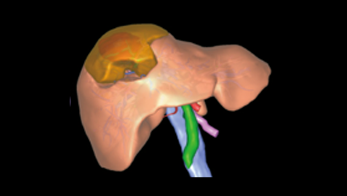
Anatomic liver segments labelling
AI technology to automatically extract liver vessels and then labels liver according to the anatomic liver segments (Couinaud segments) based on the vessel running.
This can be used for Interventional Radiology planning for liver.
This can be used for Interventional Radiology planning for liver.
Anatomy SegmentationCTITRadiologyGastroenterology
Liver analysis
Technology that automatically extracts the liver parenchyma and surrounding vessels from CT images and analyzes them in 3D.
This technology can assist preoperative simulation by planning resection area based on the calculation of the dominant vessels.
This technology can assist preoperative simulation by planning resection area based on the calculation of the dominant vessels.
Anatomy SegmentationCTITRadiologyGastroenterology
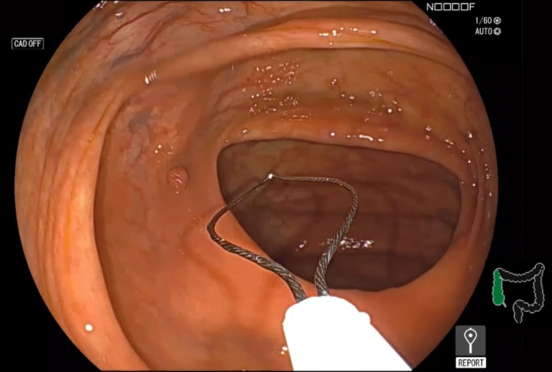
Detection assist technology for colonic polyps
This technology assists real time detection and characterization of colonic polyps from colonoscopy images with AI software.
Detection/DiagnosisESGastroenterology
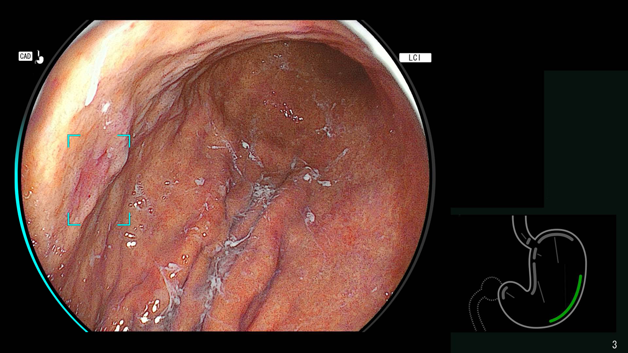
Detection technology for gastric neoplastic lesions and suspected esophageal squamous cell carcinoma
The technology utilizes AI to analyze upper gastrointestinal endoscopic images, recognizing areas suspected of being gastric neoplastic lesions or esophageal squamous cell carcinoma and detecting them in real time.
Detection/DiagnosisESGastroenterology
Endoscopy report creation support technology
In lower gastrointestinal endoscopy, AI technology is used to automatically recognize the insertion and removal of the scope from the body, as well as the insertion of surgical instruments, through software.
This is expected to support the recording of examination times and information on specimen collection (e.g., polyps), thereby reducing the burden on physicians in preparing reports.
This is expected to support the recording of examination times and information on specimen collection (e.g., polyps), thereby reducing the burden on physicians in preparing reports.
Workflow SupportESGastroenterology
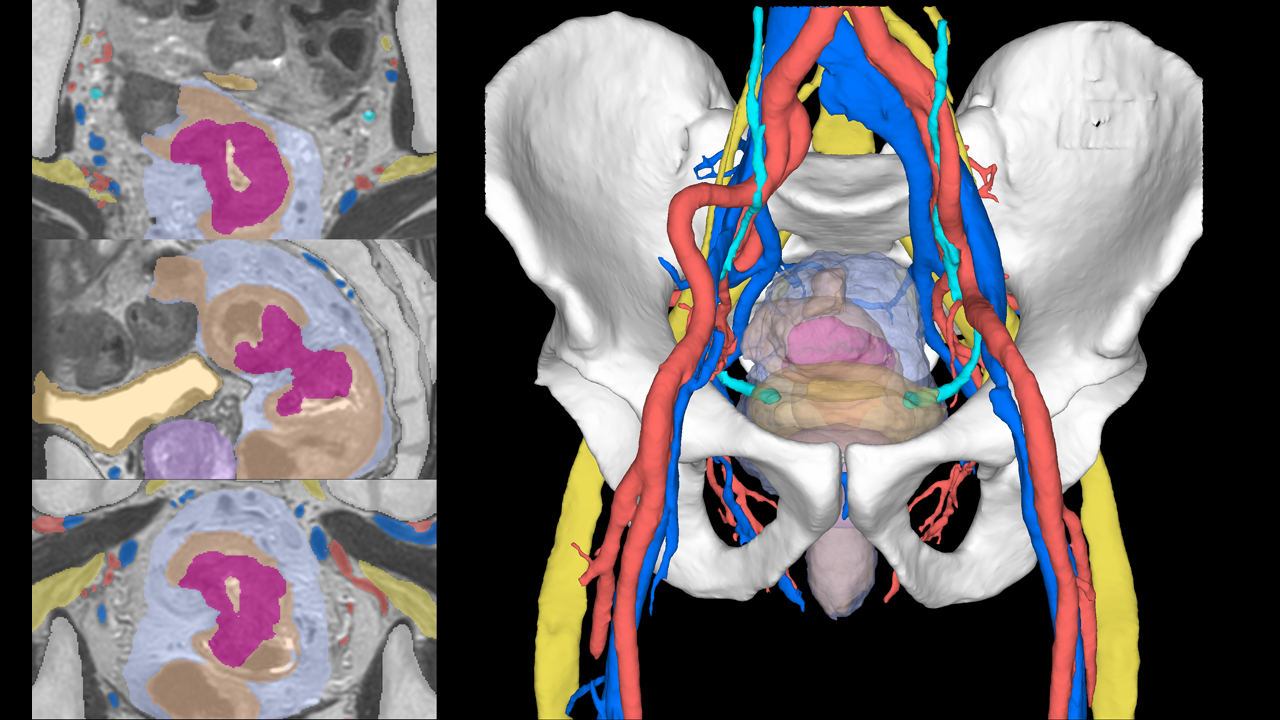
Pelvic segmentation technology
The technology for segmenting the rectum and surrounding organs from MRI images, which is expected to be useful for surgical simulation in the lower digestive tract area.
Anatomy SegmentationMRITRadiologyGastroenterology
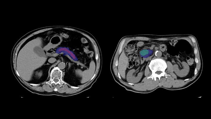
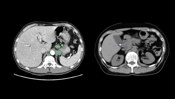
Pancreatic cancer detection technology
The technology that utilizes AI to help detect findings suggestive of pancreatic cancer from abdominal CT images.
Detection/DiagnosisITRadiologyGastroenterology
AI technology for abdominal CT
The technology that highlights areas of high/low absorption in the adrenal glands, pancreas, gallbladder, and lymph nodes compared to surrounding tissues.
Anatomy SegmentationCTITRadiologyGastroenterology
Liver deformation technology
Observe the liver and surrounding organs while deforming them, enabling estimation of the position of blood vessels during surgical detachment.
Anatomy SegmentationITRadiologyGastroenterology
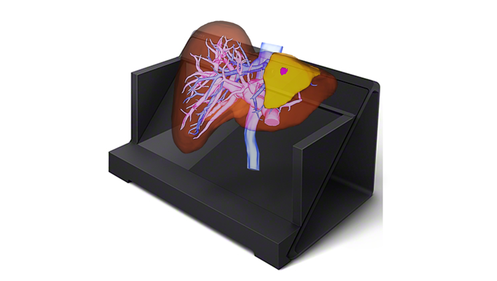
Naked-eye stereoscopic technology
Display 3D images created in the target application on a spatial reproduction display, and operate them in synchronization with our 3D system.
Anatomy SegmentationITRadiologyCardiologyRespiratoryGastroenterologyOrthopedics
AI technology for the gallbladder and pancreas
Displays the complex structure of the gallbladder and pancreas in three dimensions, supporting preoperative simulation of organ relationships and other factors.
Anatomy SegmentationITRadiologyGastroenterology
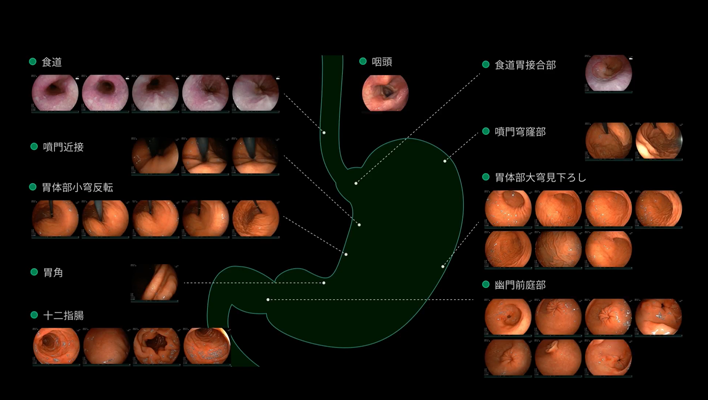
Endoscopy report creation support technology
In upper gastrointestinal endoscopy, AI technology is used to automatically recognize the location of images taken by doctors.
By extracting appropriate images for each location and automatically recording them in a report, it is expected that the burden of report creation for doctors will be reduced.
By extracting appropriate images for each location and automatically recording them in a report, it is expected that the burden of report creation for doctors will be reduced.
Workflow SupportESGastroenterology
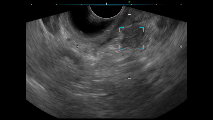
Ultrasound endoscopic diagnostic support technology for the pancreatic region
We have developed ultrasound endoscopy diagnostic support software that detects areas suspected of pancreatic solid lesions in real time during ultrasound endoscopy examinations, thereby supporting the early detection of pancreatic cancer.
By analyzing ultrasound endoscopy images, the software detects areas where the pancreas is presumed to exist and areas suspected of pancreatic solid lesions in real time, and displays the results on the monitor's ultrasound endoscopy image.
By alerting the operator, the software assists in detecting pancreatic solid lesions.
Detection/DiagnosisESRadiologyGastroenterology
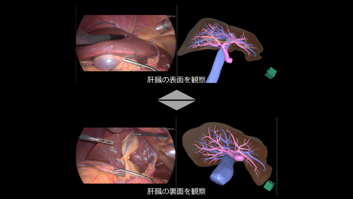
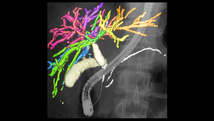
AI technology linked with 3D liver imaging
An AI technology that automatically rotates the preoperatively created 3D liver image to the same orientation as the liver in endoscopic video, allowing simultaneous reference on the same monitor, thereby supporting the understanding of internal liver structures such as blood vessels and tumor locations that are difficult to observe from the liver surface.
Workflow SupportITGastroenterology
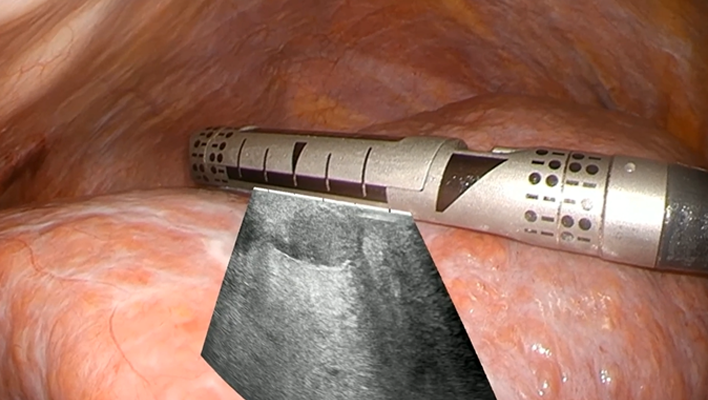
AI technology for ultrasound image overlay
An AI technology that analyzes the position and orientation of the ultrasound probe within endoscopic images during intraoperative ultrasound examinations performed in laparoscopic or robot-assisted surgeries, and overlays the ultrasound images onto the endoscopic video to support intuitive understanding of the ultrasound images.
Workflow SupportITGastroenterology