Brain segmentation
AI technology to segment and quantify the volume of each brain regions.
This technology can be used for pre-surgery simulation or calculation of the atrophy rate for each region between past and current exams.
This technology can be used for pre-surgery simulation or calculation of the atrophy rate for each region between past and current exams.
Anatomy SegmentationMRITRadiologyNeurology
Highlighting areas of higher or lower signals by comparing left/right head CT images
This technology extracts high-signal and low-signal areas in head CT images by comparing the left and right sides of the brain region. Generally, high-signal and low-signal areas are used to evaluate the state of hemorrhage and ischemia in the brain for stroke diagnosis. This will assist the diagnosis of head CT imaging.
Detection/DiagnosisCTITRadiologyNeurology
Automatic measurement of EvansIndex, corpus callosum angle, and MidlineShift
This technology automatically measures EvansIndex, corpus callosum angle, and MidlineShift from head CT. EvansIndex and corpus callosum angle are expected to support the evaluation of hydrocephalus and MidlineShift to evaluate head trauma.
Detection/DiagnosisCTITRadiologyNeurology
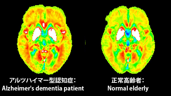
Early diagnosis of dementia
The technology under development quantitative detection of slight brain volume loss and iron deposition in the early stages of dementia using hybrid analysis of QSM(Quantitative Susceptibility Mapping)and VBM(Voxel Based Morphometry)in MRI. This will assist the diagnosis of dementia.
Detection/DiagnosisMRITRadiologyNeurology
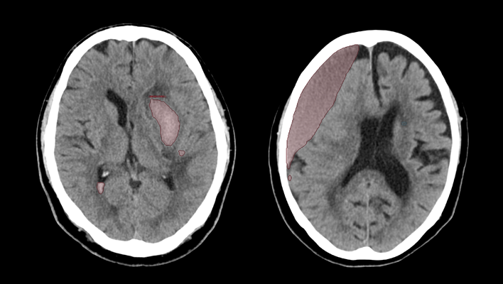
CT technology for diagnosing subarachnoid hemorrhage
We have developed AI technology to identify areas suspected of cerebral hemorrhage or cerebral infarction in CT images of the head.
This technology is expected to aid in the diagnosis of stroke by helping to evaluate bleeding and ischemia in the brain.
This technology is expected to aid in the diagnosis of stroke by helping to evaluate bleeding and ischemia in the brain.
Detection/DiagnosisCTITRadiologyNeurology
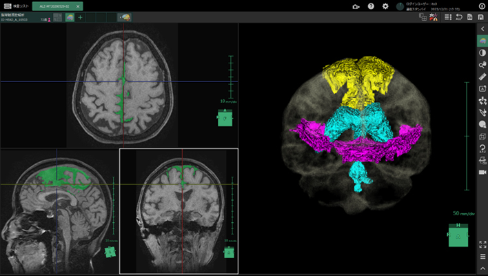
Cerebrospinal fluid analysis technology in MRI images
We have developed AI technology to extract each region of the cerebrospinal fluid cavity from MRI images.
The aim is to improve diagnostic accuracy for Hakim's disease (iNPH), a form of dementia that is important to detect early for treatment.
This technology is expected to improve the objectivity and accuracy of diagnosis by enabling AI to efficiently analyze regions associated with key findings (DESH) and support differentiation from brain atrophy.
Anatomy SegmentationMRITRadiologyNeurology
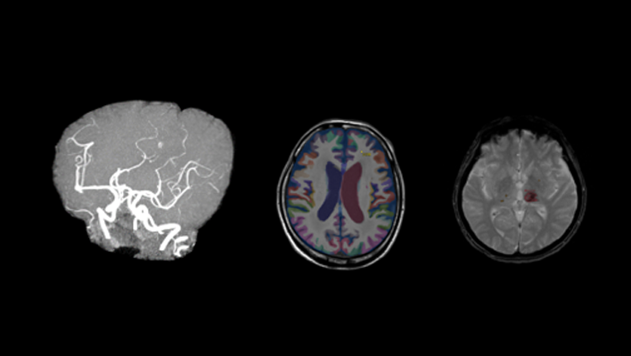
AI technology for head MRI
High signal/low signal region extraction technology, brain region labeling technology, brain extraction technology.
Anatomy SegmentationMRITRadiologyNeurology