Iterative processing utilizing AI technology to enhance visibility for low dose CT images
The technology controls image quality based on a statistical model, an object model and a physical model using iterative processing.
This technology provides a high visibility image even at a low radiation dose , by maintaining the original texture of the image at a high denoise condition.
It brings both “high visibility at low dose” and “original texture at high noise reduction”
This technology provides a high visibility image even at a low radiation dose , by maintaining the original texture of the image at a high denoise condition.
It brings both “high visibility at low dose” and “original texture at high noise reduction”
Image EnhancementCTITRadiologyCardiologyRespiratoryOrthopedics
Virtual thin slice generation technology
The technology virtually generates thin slices from thick slices. It can be applied to the whole body, useful for utilizing past data. It brings high visibility in the VR display and reconstructed sagittal/coronal images.
Image EnhancementCTITRadiologyCardiologyRespiratoryGastroenterologyOrthopedics
Lung analysis
AI technology to automatically extract lung fields, 5 lobes, and bronchus, surrounding vessels from CT images.
From each extraction result, the percentage of low attenuation area is calculated.
These are expected to contribute the diagnosis of COPD.
From each extraction result, the percentage of low attenuation area is calculated.
These are expected to contribute the diagnosis of COPD.
Anatomy SegmentationCTITRadiologyRespiratory
Lung labelling
AI technology to subdivide lung into 10 segments in right lung, 8 segments in left lung from CT images. It can be used for confirming the position of chest nodules detected by a doctor.
Anatomy SegmentationCTITRadiologyRespiratory
COVID CAD
AI technology to identify suspicious region with COVID-19 related findings from CT images. This technology will help doctors diagnose efficiently.
Detection/DiagnosisCTITRadiologyRespiratory
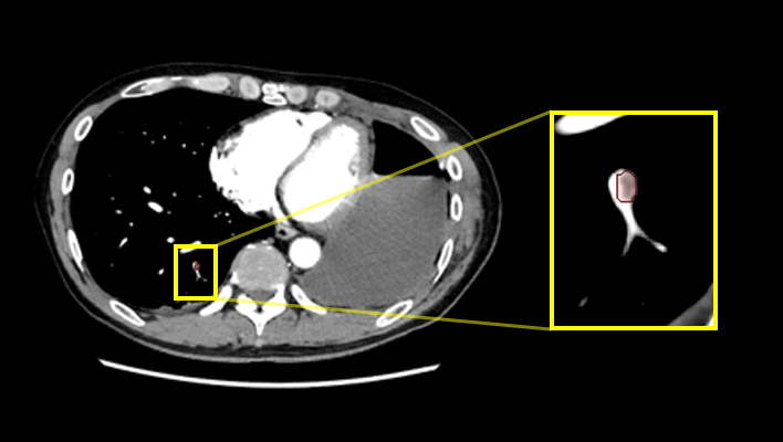
Lung nodule CAD
AI technology to detect and quantify suspicious lesion from CT Images.
This will assit prevention of overlooking of nodule and generation of language of findings for radiology report.
This will assit prevention of overlooking of nodule and generation of language of findings for radiology report.
Detection/DiagnosisCTITRadiologyRespiratory
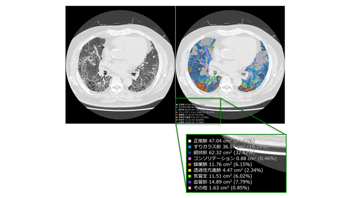
Interstitial lung disease classification
This technology identifies various findings of interstitial pneumonia that appear on CT images, such as consolidation, reticular pattern, ground glass opacity and honeycomb, and calculates their distribution and volume.
This will assist in the diagnosis of the severity and therapeutic efficacy of interstitial pneumonia, which are conventionally performed qualitatively, by providing a quantitative value for assessment.
This will assist in the diagnosis of the severity and therapeutic efficacy of interstitial pneumonia, which are conventionally performed qualitatively, by providing a quantitative value for assessment.
Detection/DiagnosisCTITRadiologyRespiratory
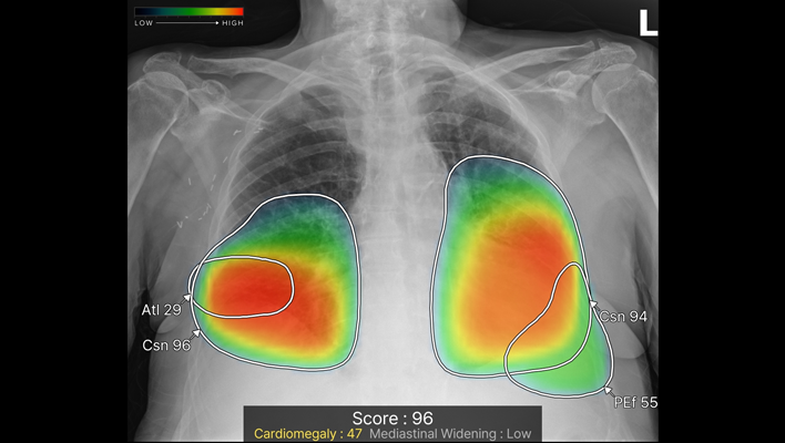
Chest X-ray CAD
The technology detects ten types of imaging findings: nodule, consolidation, pneumothorax, atelectasis, calcification, scar, pleural effusion, pneumoperitoneum, cardiomegaly and mediastinal widening from chest X-ray images. It is expected to contribute to preventing oversights in various chest X-ray examinations, such as health checkups and routine medical examinations.
Detection/DiagnosisITRadiologyRespiratory
Quantification of high absorption ROI in lung field
The technology estimates high-value threshold of the region of interest, and quantify high intensity area in lung field. For example, it is expected to offer information for quantitative analysis of partially solid nodules.
Detection/DiagnosisCTITRadiologyRespiratory
Pulmonary artery absorption enhancement technique
The technology that displays areas of low absorption in the pulmonary artery compared to surrounding tissues.
This is expected to aid in the diagnosis of pulmonary embolism.
This is expected to aid in the diagnosis of pulmonary embolism.
Anatomy SegmentationCTITRadiologyCardiologyRespiratory
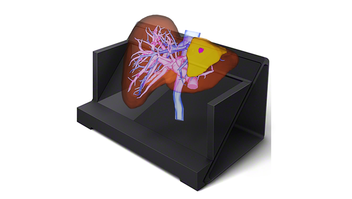
Naked-eye stereoscopic technology
Display 3D images created in the target application on a spatial reproduction display, and operate them in synchronization with our 3D system.
Anatomy SegmentationITRadiologyCardiologyRespiratoryGastroenterologyOrthopedics
AI technology for classifying the characteristics of areas suspected of interstitial lung disease and calculating their size
This AI technology identifies the anatomical structures of the lungs from chest CT images, automatically classifies characteristics based on imaging feature patterns, and simultaneously calculates their size and proportion. Additionally, to enable detailed review of the distribution of abnormalities, the lung fields are segmented by lobes, with the size and proportion of abnormalities in each region displayed.
Detection/DiagnosisITRespiratory