Iterative processing utilizing AI technology to enhance visibility for low dose CT images
The technology controls image quality based on a statistical model, an object model and a physical model using iterative processing.
This technology provides a high visibility image even at a low radiation dose , by maintaining the original texture of the image at a high denoise condition.
It brings both “high visibility at low dose” and “original texture at high noise reduction”
This technology provides a high visibility image even at a low radiation dose , by maintaining the original texture of the image at a high denoise condition.
It brings both “high visibility at low dose” and “original texture at high noise reduction”
Image EnhancementCTITRadiologyCardiologyRespiratoryOrthopedics
Denoising technique utilizing AI technology
We are developing the AI image reconstruction technology that removes noise components of image by preventing the structure and the contrast from degrading by a high speed or a high resolution MR scanning. As a result, the technology shortens the scan time with keeping the image quality, or improves the visibility of the image by improving the image quality.
Image EnhancementMRITRadiologyCardiologyGastroenterologyOrthopedics
Super resolution processing
This flitering technology with AI suppresses aliasing noises which occur in extended images.
Image EnhancementITRadiologyGastroenterology
Noise reduction technology with AI
This technology uses AI to distinguish between echo signals and noises, and extracts the signals necessary for diagnosis. This enables ultrasound systems to provide high-quality images even in difficult cases.
Image EnhancementUSRadiologyCardiologyGastroenterologyOrthopedics
Virtual thin slice generation technology
The technology virtually generates thin slices from thick slices. It can be applied to the whole body, useful for utilizing past data. It brings high visibility in the VR display and reconstructed sagittal/coronal images.
Image EnhancementCTITRadiologyCardiologyRespiratoryGastroenterologyOrthopedics
Brain segmentation
AI technology to segment and quantify the volume of each brain regions.
This technology can be used for pre-surgery simulation or calculation of the atrophy rate for each region between past and current exams.
This technology can be used for pre-surgery simulation or calculation of the atrophy rate for each region between past and current exams.
Anatomy SegmentationMRITRadiologyNeurology
Bone temporal subtraction
This technology visualizes the bone density temporal difference by performing image registration between the past and current image of the same patient.
The increase and decrease in CT value will be highlighted.
The increase and decrease in CT value will be highlighted.
Anatomy SegmentationCTITRadiologyOrthopedics
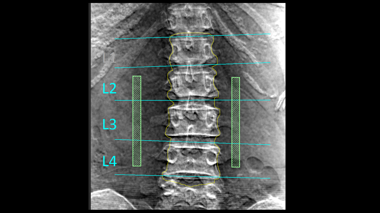
Bone Mineral Density
This technology which developed by deep learning will automatically detect the lumbar spine and femur bone that enhanced by energy subtraction, and calculate the bone mineral density.
Anatomy SegmentationDRRadiology
Pancreas analysis
Automatically extracts the pancreas, each surrounding organ area, and surrounding vessels from contrast-enhanced CT images and analyzes them in 3D.
The pancreatic duct diameter, residual pancreatic volume, measurement of the cross-sectional area, and resection simulation can be performed to support pancreatic surgery.
The pancreatic duct diameter, residual pancreatic volume, measurement of the cross-sectional area, and resection simulation can be performed to support pancreatic surgery.
Anatomy SegmentationCTITRadiologyGastroenterology
Kidney segmentation
AI technology to extract each kidney and surrounding vessels from CT or MRI images assists volume calculation of renal and compare current and past results.
This technology can also precisely extract cystic kidneys, a genetic disease that is often difficult to extract due to the diversity of shapes, and assist diagnosis and treatment.
This technology can also precisely extract cystic kidneys, a genetic disease that is often difficult to extract due to the diversity of shapes, and assist diagnosis and treatment.
Anatomy SegmentationCTMRITRadiology
Anatomic liver segments labelling
AI technology to automatically extract liver vessels and then labels liver according to the anatomic liver segments (Couinaud segments) based on the vessel running.
This can be used for Interventional Radiology planning for liver.
This can be used for Interventional Radiology planning for liver.
Anatomy SegmentationCTITRadiologyGastroenterology
Liver analysis
Technology that automatically extracts the liver parenchyma and surrounding vessels from CT images and analyzes them in 3D.
This technology can assist preoperative simulation by planning resection area based on the calculation of the dominant vessels.
This technology can assist preoperative simulation by planning resection area based on the calculation of the dominant vessels.
Anatomy SegmentationCTITRadiologyGastroenterology
Lung analysis
AI technology to automatically extract lung fields, 5 lobes, and bronchus, surrounding vessels from CT images.
From each extraction result, the percentage of low attenuation area is calculated.
These are expected to contribute the diagnosis of COPD.
From each extraction result, the percentage of low attenuation area is calculated.
These are expected to contribute the diagnosis of COPD.
Anatomy SegmentationCTITRadiologyRespiratory
Knee joint analysis
This technology automatically extracts the femur, tibia, patella, cartilage, and meniscus from MRI images.
By measuring the thickness of the cartilage and the distance between the meniscus and tibia, it is possible to evaluate the condition of the knee joint without invasion.
By measuring the thickness of the cartilage and the distance between the meniscus and tibia, it is possible to evaluate the condition of the knee joint without invasion.
Anatomy SegmentationMRITRadiologyOrthopedics
Vertebrae and ribs labelling
Technology to recognize each vertebral body individually and label the vertebrae and ribs. This reduces the burden on the physician of visually counting bone positions. It is also used for visualization of fractures and bone metastasis by subtraction of rigid body alignment of vertebral bodies.
Anatomy SegmentationCTITRadiologyOrthopedics
Lung labelling
AI technology to subdivide lung into 10 segments in right lung, 8 segments in left lung from CT images. It can be used for confirming the position of chest nodules detected by a doctor.
Anatomy SegmentationCTITRadiologyRespiratory
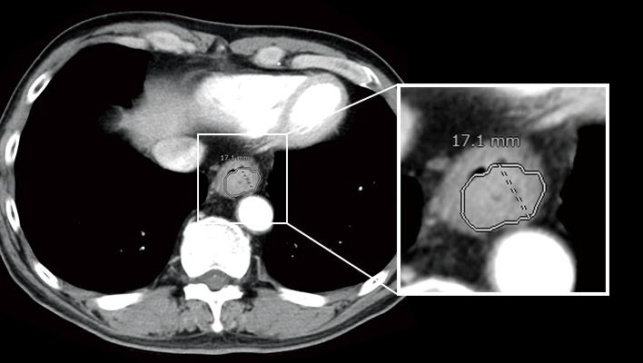
Mediastinum and axillary lymph node segmentation
AI technology to segment relatively large mediastinum, axillary lymph node from CT images. This technology can be used for assessment of lymph node enlargement.
Anatomy SegmentationCTITRadiology
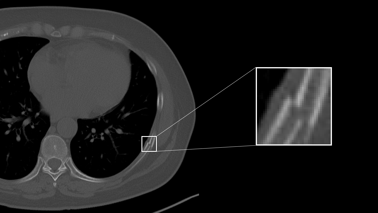
Rib fracture CAD
AI technology to detect a suspcious rib fracture from CT images. This technology will assist prevention of overlooking of subtle rib fracture.
Detection/DiagnosisCTITRadiology
COVID CAD
AI technology to identify suspicious region with COVID-19 related findings from CT images. This technology will help doctors diagnose efficiently.
Detection/DiagnosisCTITRadiologyRespiratory
Lung nodule CAD
AI technology to detect and quantify suspicious lesion from CT Images.
This will assit prevention of overlooking of nodule and generation of language of findings for radiology report.
This will assit prevention of overlooking of nodule and generation of language of findings for radiology report.
Detection/DiagnosisCTITRadiologyRespiratory
Interstitial lung disease classification
This technology identifies various findings of interstitial pneumonia that appear on CT images, such as consolidation, reticular pattern, ground glass opacity and honeycomb, and calculates their distribution and volume.
This will assist in the diagnosis of the severity and therapeutic efficacy of interstitial pneumonia, which are conventionally performed qualitatively, by providing a quantitative value for assessment.
This will assist in the diagnosis of the severity and therapeutic efficacy of interstitial pneumonia, which are conventionally performed qualitatively, by providing a quantitative value for assessment.
Detection/DiagnosisCTITRadiologyRespiratory
Highlighting areas of higher or lower signals by comparing left/right head CT images
This technology extracts high-signal and low-signal areas in head CT images by comparing the left and right sides of the brain region. Generally, high-signal and low-signal areas are used to evaluate the state of hemorrhage and ischemia in the brain for stroke diagnosis. This will assist the diagnosis of head CT imaging.
Detection/DiagnosisCTITRadiologyNeurology
Highlighting areas of higher or lower absorption than the surrounding tissue
The technology to extract and highlight areas of higher or lower absorption than the surrounding tissue. For example, high/low absorption in the liver, high absorption in the thoracic cavity, and low absorption in the kidney may be informative in determining the findings of each part. In the future, we aim to highlight both contrast-enhanced and non-contrast-enhanced images.
Detection/DiagnosisCTITRadiology
Chest X-ray CAD
The technology detects three types of imaging findings: nodule, consolidation, and pneumothorax from chest X-ray images. It is expected to contribute to preventing oversights in various chest X-ray examinations, such as health checkups and routine medical examinations.
Detection/DiagnosisITRadiologyRespiratory
Real-time Screening Assist
This technology highlights areas in the B-mode image where luminance characteristics are different from the surroundings
Detection/DiagnosisUSRadiology
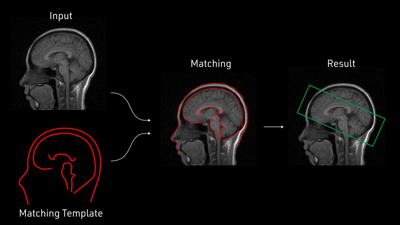
Improvement of workflow with AI
This technology extracts the target area from the image.
Positioning during imaging is performed automatically.
Unnecessary tissue is automatically removed before creating a 3D image for diagnosis.
It is a technology that supports the automation of inspections.
Positioning during imaging is performed automatically.
Unnecessary tissue is automatically removed before creating a 3D image for diagnosis.
It is a technology that supports the automation of inspections.
Workflow SupportMRITRadiology
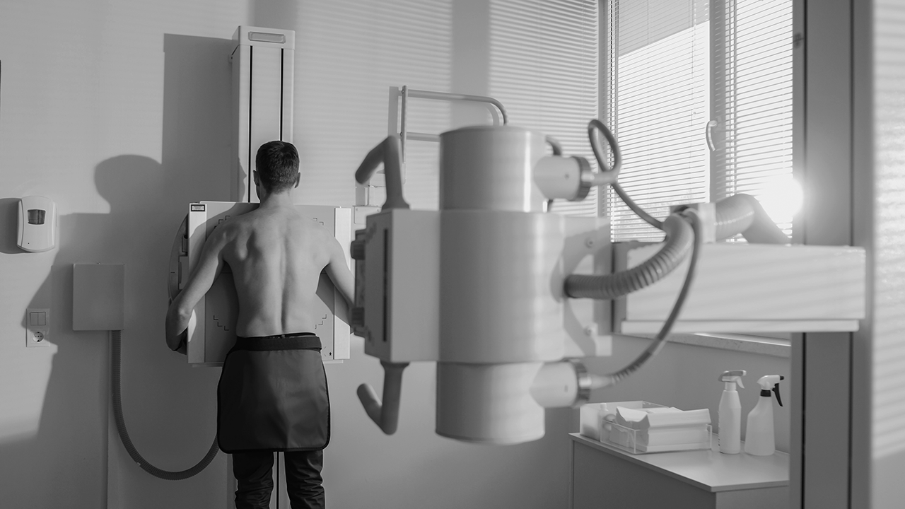
Positioning assist
This technology is to assist positioning before exposure by images from camera which implemented on the X-ray modality and confirming the more optimal positioin with AI software .
Workflow SupportDRRadiology
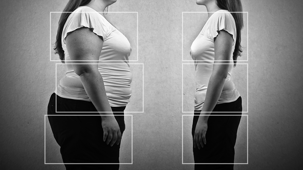
Exposure condition navigation
The sensor integrated in the X-ray system will recognize three-dimensional information from the patient's body.
It will then automatically suggest the optimized exposure conditions and adapt proper image processing.
It will then automatically suggest the optimized exposure conditions and adapt proper image processing.
Workflow SupportDRRadiology
Lung nodule characterization analysis
The technology supports populating radiology report of lung nodules based on the observation and analysis results of the properties. It enables to analyze multiple properties such as shape, absorption, presence of calcification, etc., and provide multiple finding sentence candidates using natural language processing technology.
Workflow SupportCTITRadiology
Liver characterization analysis
The technology analyzes the properties of areas of interest in the liver and assists doctors in writing radiology report. It is possible to analyze not only the location and size, but also the appearance/disappearance of enhancement based on multi-phase images of contrast-enhanced CT. In addition, similar to lung nodule characterization analysis, it is also possible to present multiple finding sentence candidates using natural language processing technology.
Workflow SupportCTITRadiology
Quantification of high absorption ROI in lung field
The technology estimates high-value threshold of the region of interest, and quantify high intensity area in lung field. For example, it is expected to offer information for quantitative analysis of partially solid nodules.
Detection/DiagnosisCTITRadiologyRespiratory
Automatic measurement of EvansIndex, corpus callosum angle, and MidlineShift
This technology automatically measures EvansIndex, corpus callosum angle, and MidlineShift from head CT. EvansIndex and corpus callosum angle are expected to support the evaluation of hydrocephalus and MidlineShift to evaluate head trauma.
Detection/DiagnosisCTITRadiologyNeurology
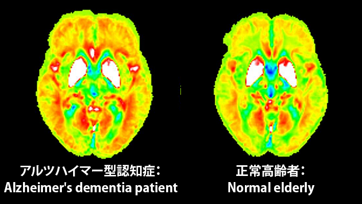
Early diagnosis of dementia
The technology under development quantitative detection of slight brain volume loss and iron deposition in the early stages of dementia using hybrid analysis of QSM(Quantitative Susceptibility Mapping)and VBM(Voxel Based Morphometry)in MRI. This will assist the diagnosis of dementia.
Detection/DiagnosisMRITRadiologyNeurology
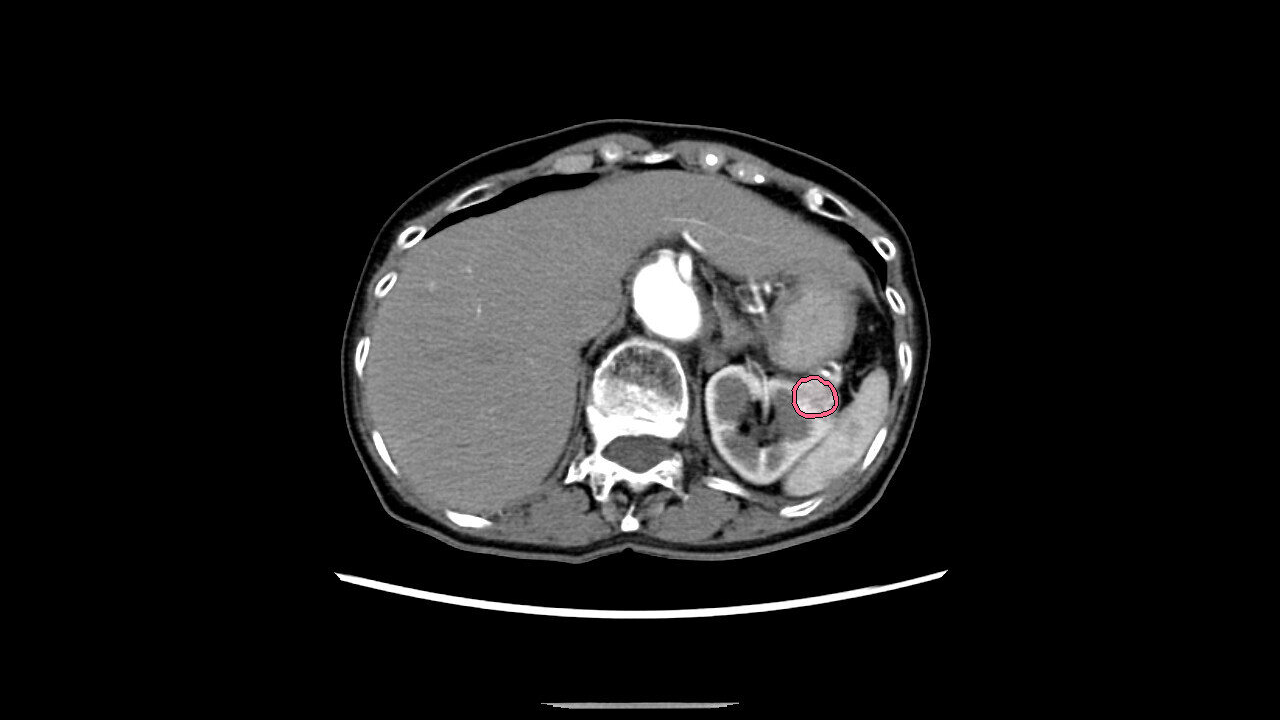
AI technology for abdominal CT
Technology that displays areas of high and low absorption in the liver, kidneys, and spleen compared to surrounding tissues.
Anatomy SegmentationCTITRadiology
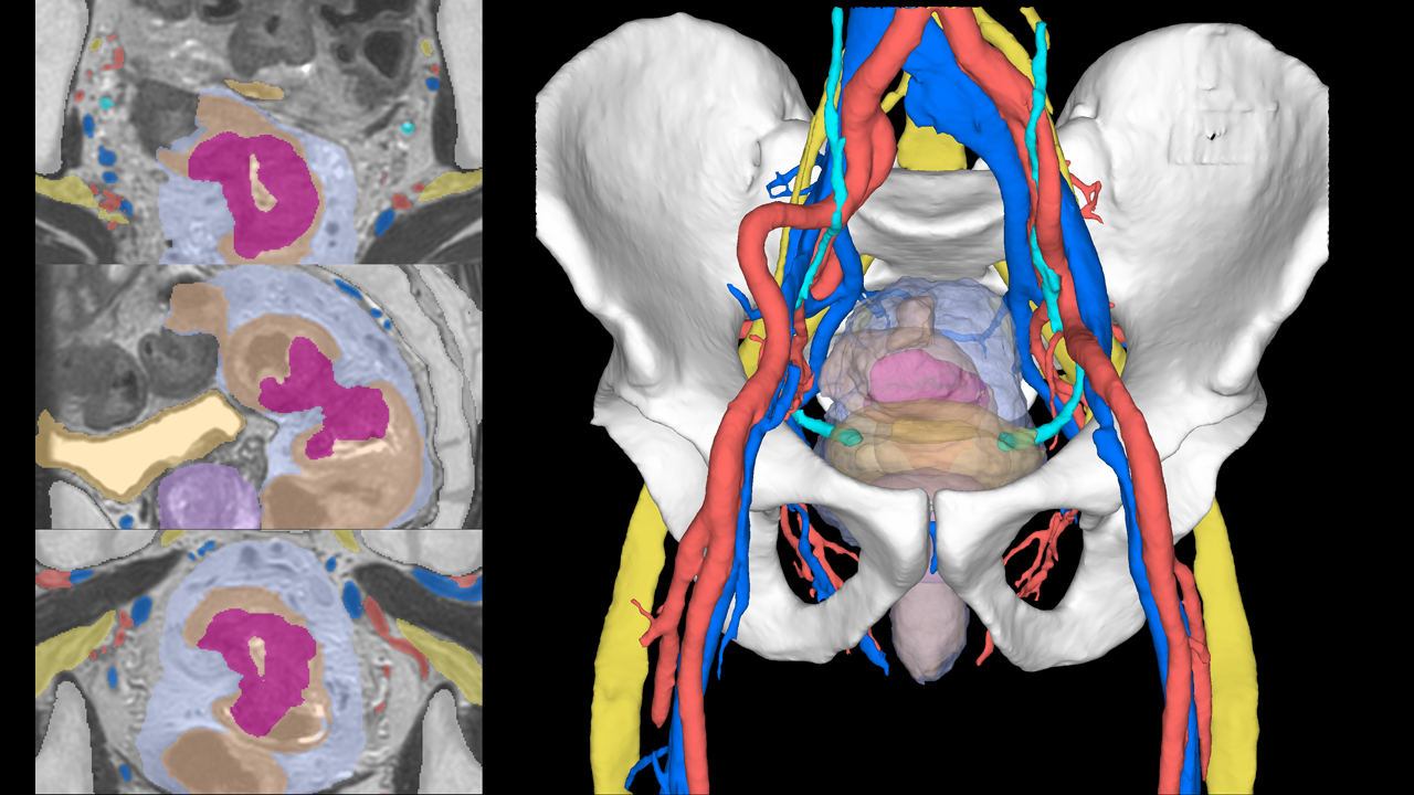
Pelvic segmentation technology
The technology for segmenting the rectum and surrounding organs from MRI images, which is expected to be useful for surgical simulation in the lower digestive tract area.
Anatomy SegmentationMRITRadiologyGastroenterology
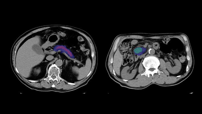
Pancreatic cancer detection technology
The technology that utilizes AI to help detect findings suggestive of pancreatic cancer from abdominal CT images.
Detection/DiagnosisITRadiologyGastroenterology
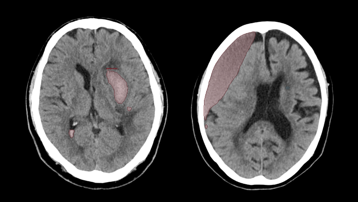
CT technology for diagnosing subarachnoid hemorrhage
We have developed AI technology to identify areas suspected of cerebral hemorrhage or cerebral infarction in CT images of the head.
This technology is expected to aid in the diagnosis of stroke by helping to evaluate bleeding and ischemia in the brain.
This technology is expected to aid in the diagnosis of stroke by helping to evaluate bleeding and ischemia in the brain.
Detection/DiagnosisCTITRadiologyNeurology
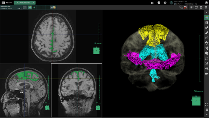
Cerebrospinal fluid analysis technology in MRI images
We have developed AI technology to extract each region of the cerebrospinal fluid cavity from MRI images.
The aim is to improve diagnostic accuracy for Hakim's disease (iNPH), a form of dementia that is important to detect early for treatment.
This technology is expected to improve the objectivity and accuracy of diagnosis by enabling AI to efficiently analyze regions associated with key findings (DESH) and support differentiation from brain atrophy.
Anatomy SegmentationMRITRadiologyNeurology
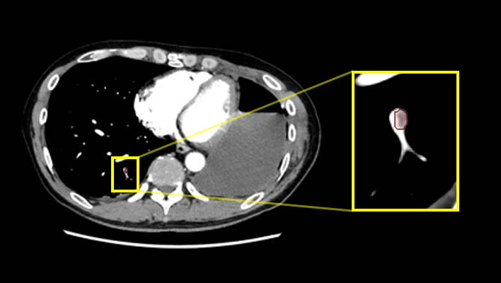
Pulmonary artery absorption enhancement technique
The technology that displays areas of low absorption in the pulmonary artery compared to surrounding tissues.
This is expected to aid in the diagnosis of pulmonary embolism.
This is expected to aid in the diagnosis of pulmonary embolism.
Anatomy SegmentationCTITRadiologyCardiologyRespiratory
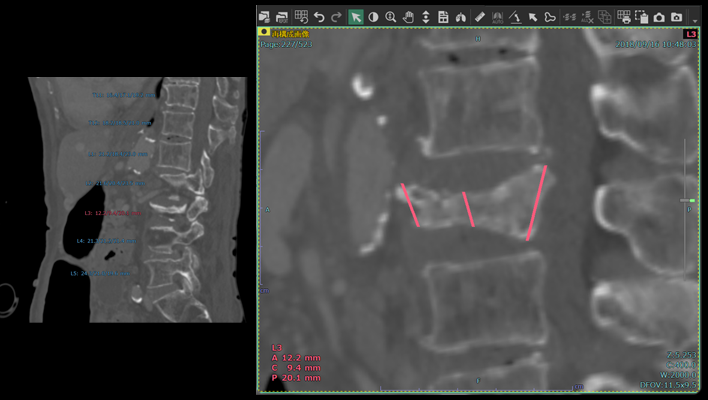
Automatic vertebral body height measurement technology
The technology that automatically measures the height of each vertebral body and displays the results classified according to user-defined thresholds.
This technology is expected to aid in the diagnosis of vertebral fractures.
This technology is expected to aid in the diagnosis of vertebral fractures.
Anatomy SegmentationITRadiologyOrthopedics
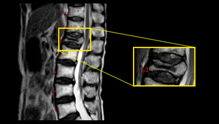
MRI labeling technology
The technique for labeling vertebral numbers on MRI images.
Anatomy SegmentationMRITRadiologyOrthopedics
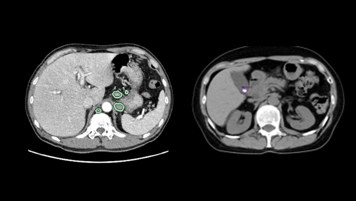
AI technology for abdominal CT
The technology that highlights areas of high/low absorption in the adrenal glands, pancreas, gallbladder, and lymph nodes compared to surrounding tissues.
Anatomy SegmentationCTITRadiologyGastroenterology
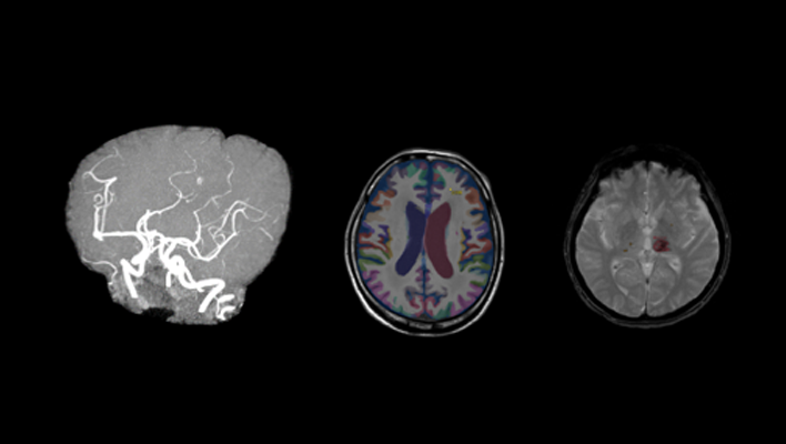
AI technology for head MRI
High signal/low signal region extraction technology, brain region labeling technology, brain extraction technology.
Anatomy SegmentationMRITRadiologyNeurology
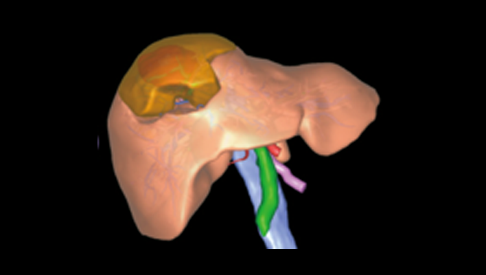
Liver deformation technology
Observe the liver and surrounding organs while deforming them, enabling estimation of the position of blood vessels during surgical detachment.
Anatomy SegmentationITRadiologyGastroenterology
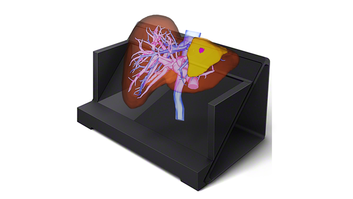
Naked-eye stereoscopic technology
Display 3D images created in the target application on a spatial reproduction display, and operate them in synchronization with our 3D system.
Anatomy SegmentationITRadiologyCardiologyRespiratoryGastroenterologyOrthopedics
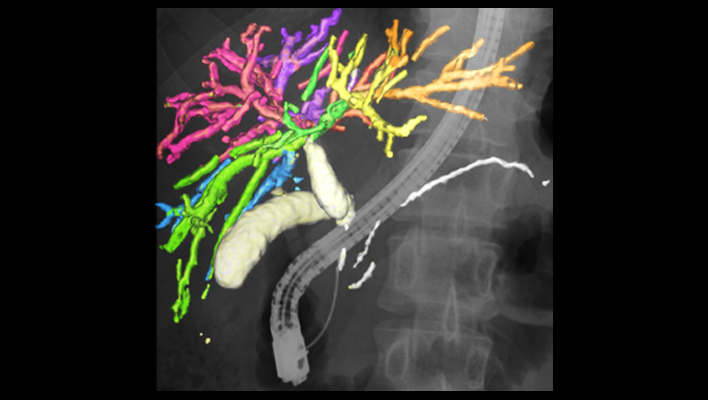
AI technology for the gallbladder and pancreas
Displays the complex structure of the gallbladder and pancreas in three dimensions, supporting preoperative simulation of organ relationships and other factors.
Anatomy SegmentationITRadiologyGastroenterology
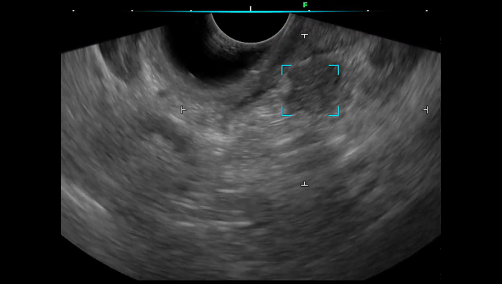
Ultrasound endoscopic diagnostic support technology for the pancreatic region
We have developed ultrasound endoscopy diagnostic support software that detects areas suspected of pancreatic solid lesions in real time during ultrasound endoscopy examinations, thereby supporting the early detection of pancreatic cancer.
By analyzing ultrasound endoscopy images, the software detects areas where the pancreas is presumed to exist and areas suspected of pancreatic solid lesions in real time, and displays the results on the monitor's ultrasound endoscopy image.
By alerting the operator, the software assists in detecting pancreatic solid lesions.
Detection/DiagnosisESRadiologyGastroenterology