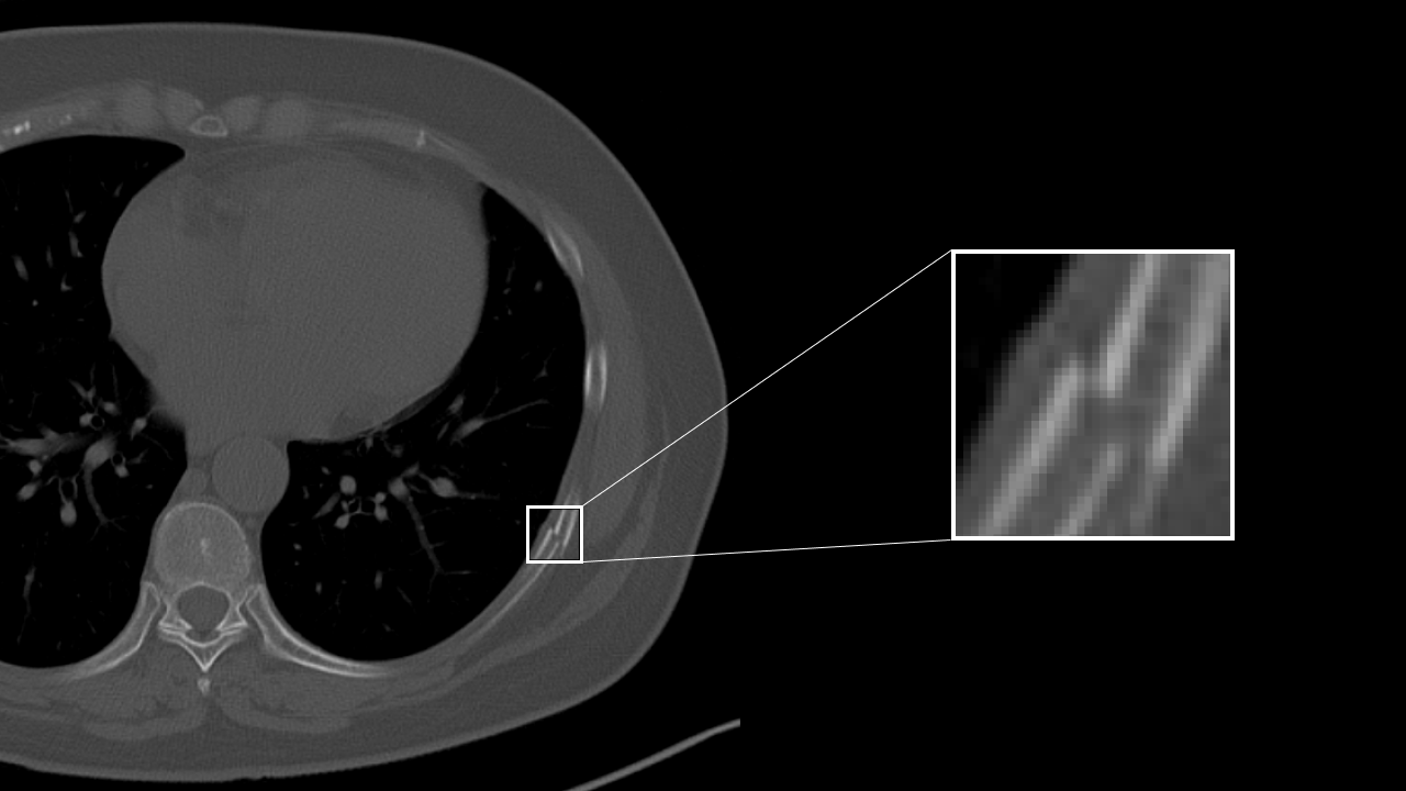
Rib fracture CAD
AI technology to detect a suspcious rib fracture from CT images. This technology will assist prevention of overlooking of subtle rib fracture.
Detection/DiagnosisCTITRadiology
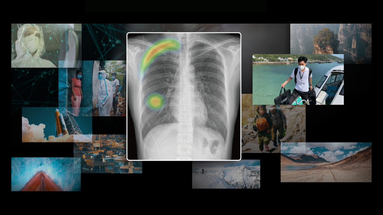
COVID CAD
AI technology to identify suspicious region with COVID-19 related findings from CT images. This technology will help doctors diagnose efficiently.
Detection/DiagnosisCTITRadiologyRespiratory
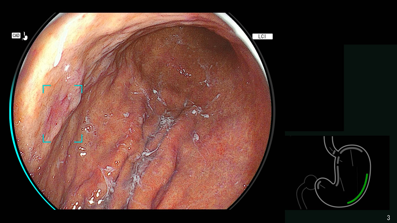
Detection assist technology for colonic polyps
This technology assists real time detection and characterization of colonic polyps from colonoscopy images with AI software.
Detection/DiagnosisESGastroenterology
Lung nodule CAD
AI technology to detect and quantify suspicious lesion from CT Images.
This will assit prevention of overlooking of nodule and generation of language of findings for radiology report.
This will assit prevention of overlooking of nodule and generation of language of findings for radiology report.
Detection/DiagnosisCTITRadiologyRespiratory
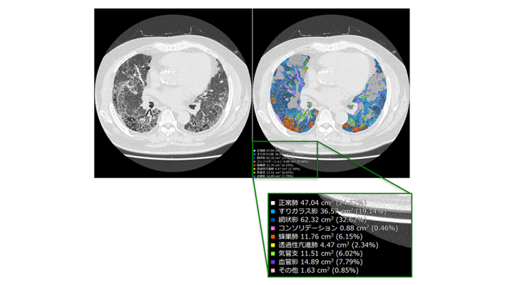
Interstitial lung disease classification
This technology identifies various findings of interstitial pneumonia that appear on CT images, such as consolidation, reticular pattern, ground glass opacity and honeycomb, and calculates their distribution and volume.
This will assist in the diagnosis of the severity and therapeutic efficacy of interstitial pneumonia, which are conventionally performed qualitatively, by providing a quantitative value for assessment.
This will assist in the diagnosis of the severity and therapeutic efficacy of interstitial pneumonia, which are conventionally performed qualitatively, by providing a quantitative value for assessment.
Detection/DiagnosisCTITRadiologyRespiratory
AI-CAD in modality
The technology which prepares environment on the modality to exapnd the opportuities of AI-CAD applications and improve workflow not only in hospitals, but also at point of care.
Detection/DiagnosisDRothers
Highlighting areas of higher or lower signals by comparing left/right head CT images
This technology extracts high-signal and low-signal areas in head CT images by comparing the left and right sides of the brain region. Generally, high-signal and low-signal areas are used to evaluate the state of hemorrhage and ischemia in the brain for stroke diagnosis. This will assist the diagnosis of head CT imaging.
Detection/DiagnosisCTITRadiologyNeurology
Highlighting areas of higher or lower absorption than the surrounding tissue
The technology to extract and highlight areas of higher or lower absorption than the surrounding tissue. For example, high/low absorption in the liver, high absorption in the thoracic cavity, and low absorption in the kidney may be informative in determining the findings of each part. In the future, we aim to highlight both contrast-enhanced and non-contrast-enhanced images.
Detection/DiagnosisCTITRadiology
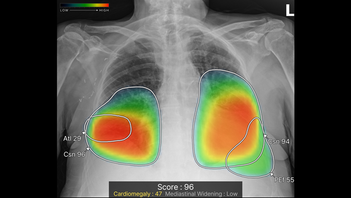
Chest X-ray CAD
The technology detects ten types of imaging findings: nodule, consolidation, pneumothorax, atelectasis, calcification, scar, pleural effusion, pneumoperitoneum, cardiomegaly and mediastinal widening from chest X-ray images. It is expected to contribute to preventing oversights in various chest X-ray examinations, such as health checkups and routine medical examinations.
Detection/DiagnosisITRadiologyRespiratory
Real-time Screening Assist
This technology highlights areas in the B-mode image where luminance characteristics are different from the surroundings
Detection/DiagnosisUSRadiology
Quantification of high absorption ROI in lung field
The technology estimates high-value threshold of the region of interest, and quantify high intensity area in lung field. For example, it is expected to offer information for quantitative analysis of partially solid nodules.
Detection/DiagnosisCTITRadiologyRespiratory
Automatic measurement of EvansIndex, corpus callosum angle, and MidlineShift
This technology automatically measures EvansIndex, corpus callosum angle, and MidlineShift from head CT. EvansIndex and corpus callosum angle are expected to support the evaluation of hydrocephalus and MidlineShift to evaluate head trauma.
Detection/DiagnosisCTITRadiologyNeurology
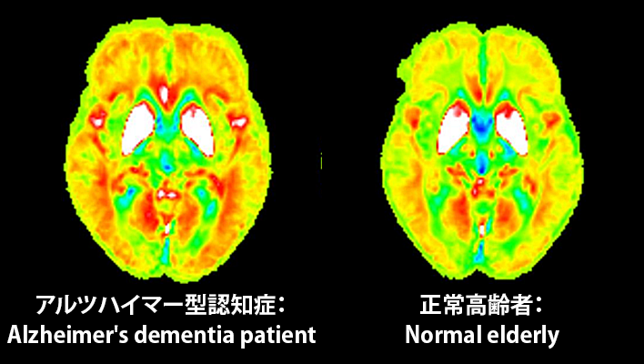
Early diagnosis of dementia
The technology under development quantitative detection of slight brain volume loss and iron deposition in the early stages of dementia using hybrid analysis of QSM(Quantitative Susceptibility Mapping)and VBM(Voxel Based Morphometry)in MRI. This will assist the diagnosis of dementia.
Detection/DiagnosisMRITRadiologyNeurology
Detection technology for gastric neoplastic lesions and suspected esophageal squamous cell carcinoma
The technology utilizes AI to analyze upper gastrointestinal endoscopic images, recognizing areas suspected of being gastric neoplastic lesions or esophageal squamous cell carcinoma and detecting them in real time.
Detection/DiagnosisESGastroenterology
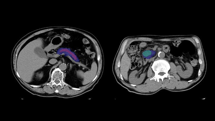
Pancreatic cancer detection technology
The technology that utilizes AI to help detect findings suggestive of pancreatic cancer from abdominal CT images.
Detection/DiagnosisITRadiologyGastroenterology
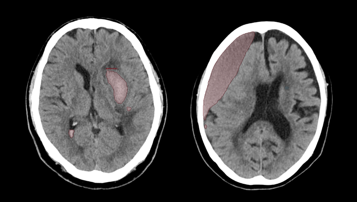
CT technology for diagnosing subarachnoid hemorrhage
We have developed AI technology to identify areas suspected of cerebral hemorrhage or cerebral infarction in CT images of the head.
This technology is expected to aid in the diagnosis of stroke by helping to evaluate bleeding and ischemia in the brain.
This technology is expected to aid in the diagnosis of stroke by helping to evaluate bleeding and ischemia in the brain.
Detection/DiagnosisCTITRadiologyNeurology
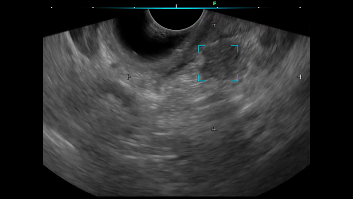
Ultrasound endoscopic diagnostic support technology for the pancreatic region
We have developed ultrasound endoscopy diagnostic support software that detects areas suspected of pancreatic solid lesions in real time during ultrasound endoscopy examinations, thereby supporting the early detection of pancreatic cancer.
By analyzing ultrasound endoscopy images, the software detects areas where the pancreas is presumed to exist and areas suspected of pancreatic solid lesions in real time, and displays the results on the monitor's ultrasound endoscopy image.
By alerting the operator, the software assists in detecting pancreatic solid lesions.
Detection/DiagnosisESRadiologyGastroenterology
AI technology for classifying the characteristics of areas suspected of interstitial lung disease and calculating their size
This AI technology identifies the anatomical structures of the lungs from chest CT images, automatically classifies characteristics based on imaging feature patterns, and simultaneously calculates their size and proportion. Additionally, to enable detailed review of the distribution of abnormalities, the lung fields are segmented by lobes, with the size and proportion of abnormalities in each region displayed.
Detection/DiagnosisITRespiratory