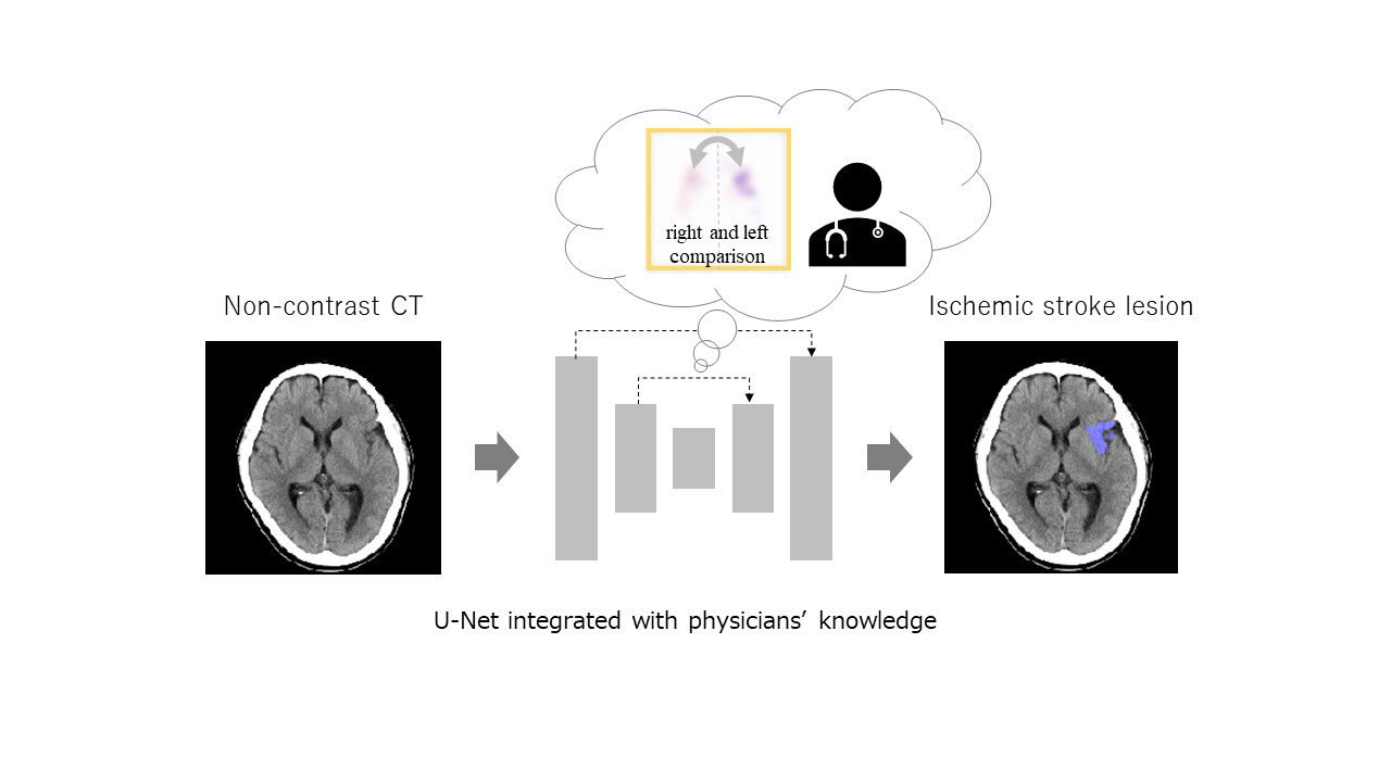
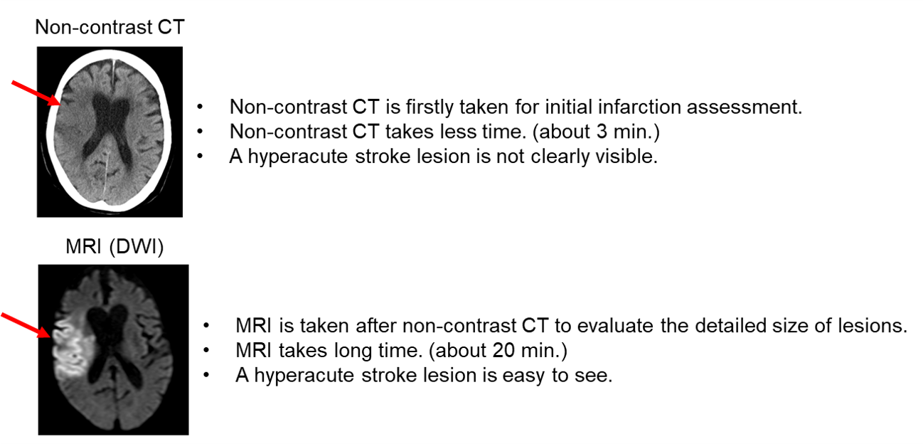
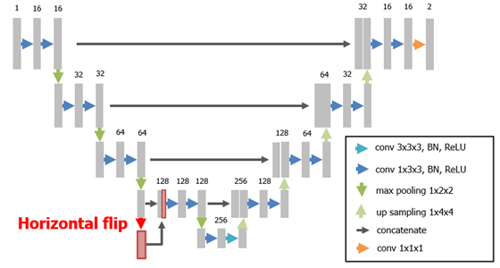
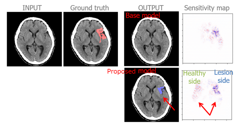
It is important to evaluate size of hyperacute stroke lesions and start therapy speedily because human nervous tissue is rapidly lost as hyperacute stroke progresses. Although it is desirable to evaluate it from non-contrast CT, which is the first choice of imaging examination for a patient with brain stroke suspicion, non-contrast CT images have only subtle CT hypoattenuation and texture variation caused by hyperacute ischemia (Figure 1). Physicians usually evaluate such images by comparing the difference between right and left sides based on the symmetric characteristic of brain anatomy not to miss the subtle lesions. In this paper [1], authors propose a convolutional neural network that integrates the comparison knowledge to automatically segment hyperacute stroke lesions (Figure 2). Experimental results showed that the proposed architecture improved segmentation accuracy (Table 1). In addition, sensitivity maps of proposed trained model demonstrated that both the right and left sides were utilized more effectively (Figure 3). In the future, this method will help to evaluate hyperacute stroke lesions more speedily and reduce time to start therapy.
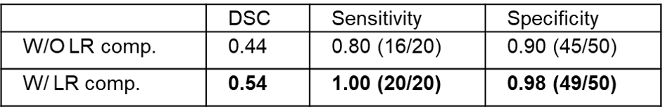
DSC: Dice similarity coefficient (the closer the value is to 1, the higher the similarity with ground truth.)
W/O LR comp.: the network without right and left comparison architecture.
W/ LR comp.: the network with right and left comparison architecture.
[1] Takuya Fuchigami, Sadato Akahori, Takayuki Okatani, and Yuanzhong Li “A hyperacute stroke segmentation method using 3D U-Net integrated with physicians’ knowledge for NCCT”, Proc. SPIE 11314, Medical Imaging 2020: Computer-Aided Diagnosis, 113140G (16 March 2020)
DOI: https://doi.org/10.1117/12.2549176
CAUTION:This is Fujifilm Global Website. Fujifilm makes no representation that products on this website are commercially available in all countries. Approved uses of products vary by country and region. Specifications and appearance of products are subject to change without notice.