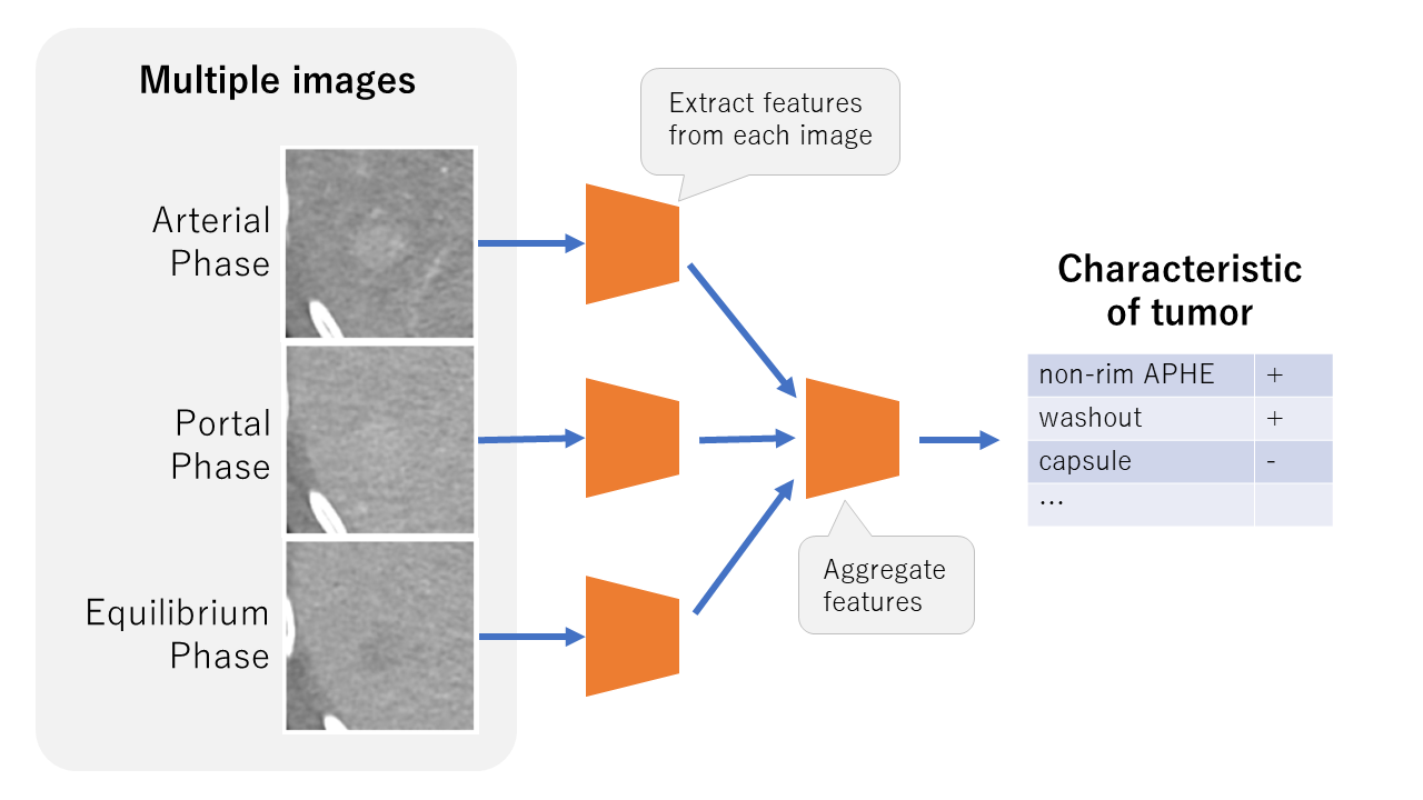
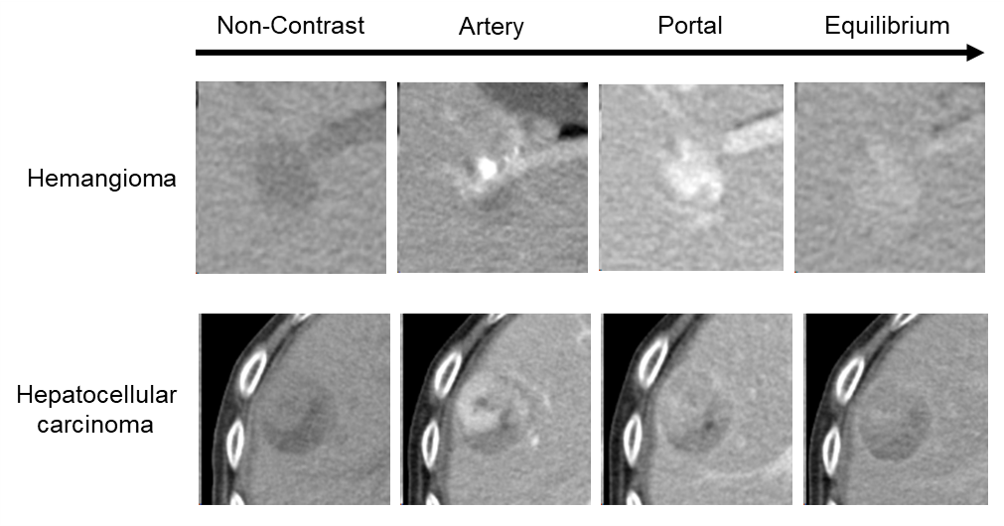
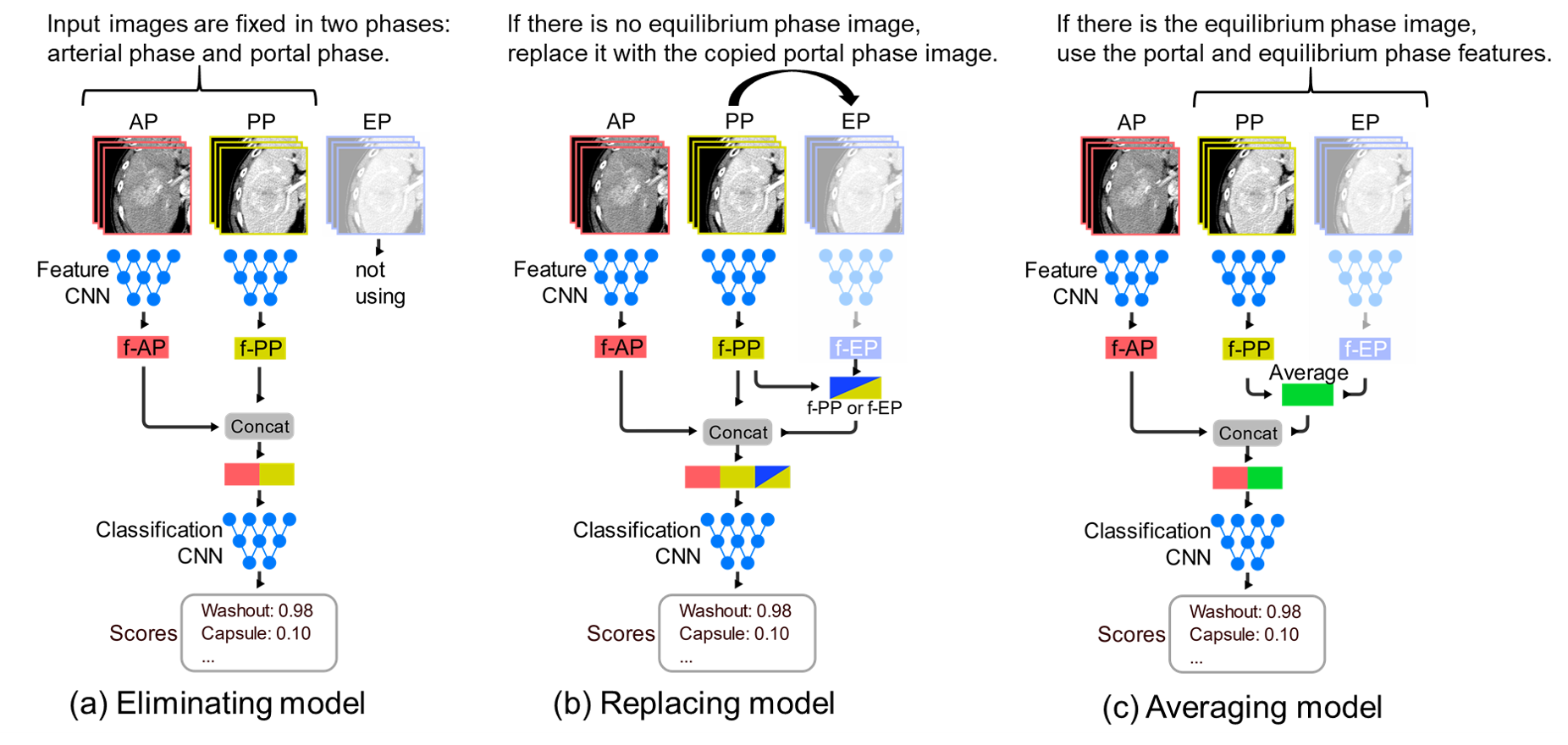
The liver is the largest solid organ in the body which performs vital functions such as creating essential nutrients and removing waste products. Distinguishing the types of liver tumors is important for determining treatment strategy, and dynamic contrast-enhanced CT (DCE-CT) provides useful characteristics for it. In this paper [1], the authors propose a deep learning neural network model for classifying tumor characteristics in DCE-CT images (Figure 2). This model is trained to classify eight features, such as non–rim APHE (non–rim arterial phase hyper enhancement) and washout, which are important for differentiating liver tumors. In addition, they propose three deep neural network classification models that can deal with variable number of input images. Evaluation results showed high discrimination performance exceeding 91% in ROC-AUC on average. In the future, it is hoped that the models will help to deal with liver tumors more accurately and quickly.
DOI: http://doi.org/10.1117/12.2648644
CAUTION:This is Fujifilm Global Website. Fujifilm makes no representation that products on this website are commercially available in all countries. Approved uses of products vary by country and region. Specifications and appearance of products are subject to change without notice.