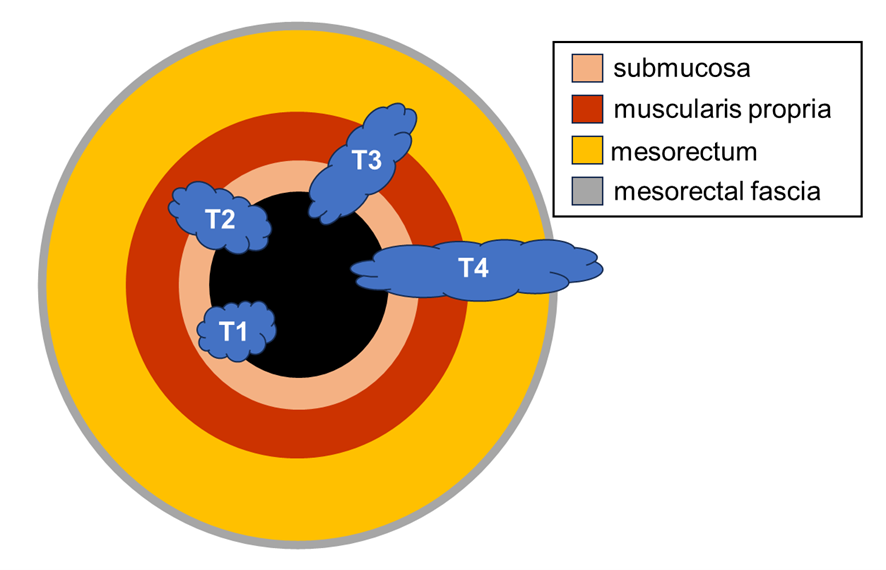
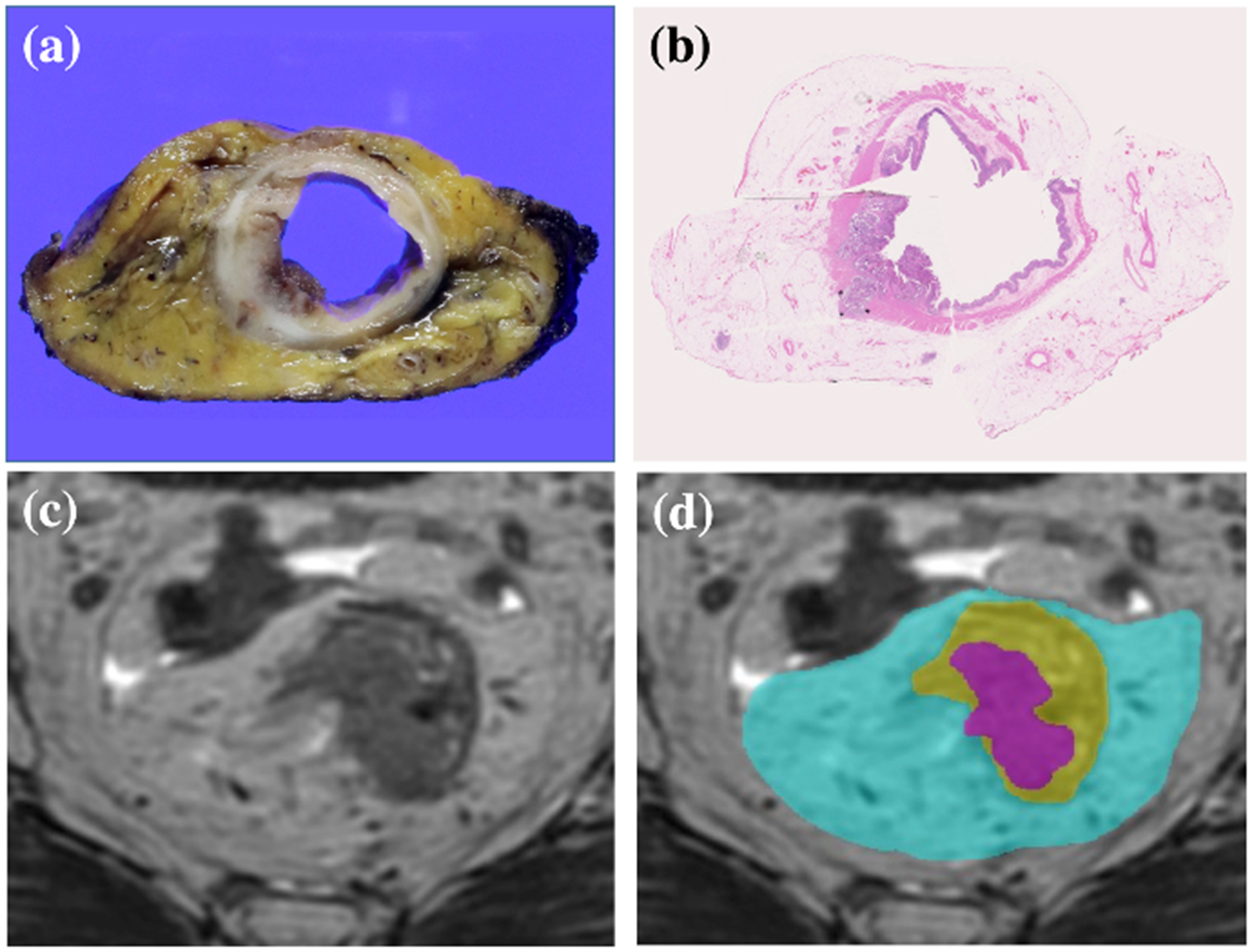
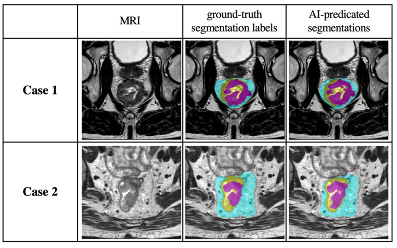
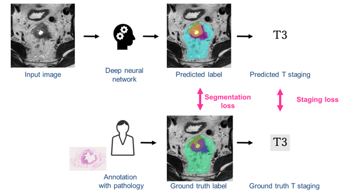
Rectal cancer is a kind of cancer that starts as a growth of cells in the rectum and as it progresses, it spreads deeper into the body (Figure 1). In order to determine the treatment strategy, it is important to extract cancer area and judge T-stage. However, it is difficult to evaluate the index from preoperative MRI images at all facilities because it requires high radiologists’ skill and experience. The paper [1] proposed the algorithm for the segmentation of the tumor, rectum, and mesorectum and staging based on them. The training data were prepared by annotating MR images referring to pathologically confirmed lesions (Figure 2). The sensitivity, specificity and accuracy of the classification for stage T2/T3 were 0.773, 0.768 and 0.771, and Dice similarity coefficients for rectal cancer, rectum, and mesorectum were 0.727, 0.930, and 0.917, respectively (Figs. 3 and 4). In the future, the system will provide stable treatment at any institution and play an important role for individualized medical care.


DOI: https://doi.org/10.1371/journal.pone.0269931
CAUTION:This is Fujifilm Global Website. Fujifilm makes no representation that products on this website are commercially available in all countries. Approved uses of products vary by country and region. Specifications and appearance of products are subject to change without notice.
