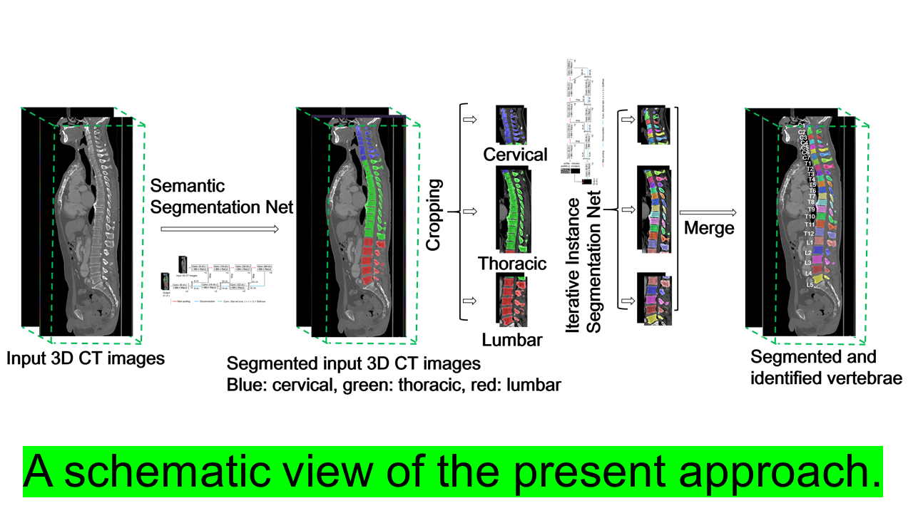
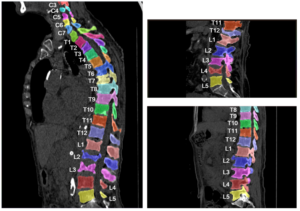
When a radiologist interprets vertebrae, it is important to identify of individual vertebrae. In automating this task, automatic segmentation, localization, and identification of individual vertebrae from 3D CT (Computed Tomography) images play an important role. In this paper[1], the authors proposed a single multi-stage framework that performs all these tasks without any assumptions. In the first stage, the cervical, thoracic, and lumbar vertebrae are detected. In the second stage, individual vertebrae are segmented and identified using the first stage’s results. The proposed method is evaluated in terms of segmentation, localization, and identification accuracy, and achieved better performance than all existing studies on all three metrics. In the future, the authors hope that the proposed method will help doctors in clinical practice.

DOI: https://doi.org/10.48550/arXiv.2009.13798
CAUTION:This is Fujifilm Global Website. Fujifilm makes no representation that products on this website are commercially available in all countries. Approved uses of products vary by country and region. Specifications and appearance of products are subject to change without notice.